Anatomy of the Temporal and Infratemporal Fossae - kapak
Bilim#temporal fossa#infratemporal fossa#anatomy#skull

Anatomy of the Temporal and Infratemporal Fossae

Explore the detailed anatomy of the temporal and infratemporal fossae, including their boundaries, contents, and critical clinical significance for head and facial regions.

selbu0909January 30, 2026 ~24 dk toplam
01

Sesli Özet

9 dakika

Konuyu otobüste, koşarken, yolda dinleyerek öğren.

Sesli Özet

Anatomy of the Temporal and Infratemporal Fossae

0:009:23
02

Flash Kartlar

25 kart

Karta tıklayarak çevir. ← → ile gez, ⎵ ile çevir.

1 / 25
Tüm kartları metin olarak gör
  1. 1. Define the temporal fossa and state its general location.

    The temporal fossa is a depressed area situated on each side of the skull, specifically within the temporal region. Its location is between the superior and inferior temporal lines and the zygomatic arch. Crucially, it is positioned superiorly to the infratemporal fossa, establishing a clear anatomical relationship between these two regions.

  2. 2. What are the postero-superior and inferior boundaries of the temporal fossa?

    Postero-superiorly, the limit of the temporal fossa is marked by the superior temporal line. Inferiorly, it is bounded by the infratemporal crest. These boundaries help delineate the extent of this important anatomical region.

  3. 3. Describe the anterior and lateral boundaries of the temporal fossa.

    Anteriorly, the boundary of the temporal fossa is formed by the frontal process of the zygomatic bone. Laterally, the prominent zygomatic arch defines its extent. These structures provide clear anatomical landmarks for the fossa.

  4. 4. Which four bones form the floor of the temporal fossa?

    The floor of the temporal fossa is a composite structure, formed by the articulation of four distinct bones. These bones are the frontal, parietal, temporal, and sphenoid bones. Their articulation creates a crucial base for the fossa's contents.

  5. 5. What is the pterion, and why is it clinically significant?

    The pterion is an important anatomical landmark within the floor of the temporal fossa, renowned as the thinnest part of the lateral wall of the skull. It is the point where the anteroinferior corner of the parietal bone articulates with the greater wing of the sphenoid bone. Clinically, it directly overlies the anterior division of the middle meningeal artery, making it vulnerable to severe intracranial hemorrhage upon trauma.

  6. 6. What is the primary muscle located within the temporal fossa?

    The primary occupant of the temporal fossa is the temporalis muscle. This is a fan-shaped muscle that plays a key role in mastication. It is one of the most powerful muscles involved in jaw movement.

  7. 7. Describe the origin and insertion of the temporalis muscle.

    The temporalis muscle originates from the superior temporal line. Its fibers converge inferiorly to form a strong tendon, which then attaches to the anterior surface of the coronoid process of the mandible. This attachment allows it to exert significant force on the jaw.

  8. 8. What is the main function of the temporalis muscle?

    The temporalis muscle is a powerful elevator and retractor of the mandible. It plays a key role in mastication, or chewing, by lifting the lower jaw and pulling it backward. This action is essential for biting and grinding food.

  9. 9. How is the temporalis muscle innervated and supplied with blood?

    The temporalis muscle is innervated by the deep temporal nerves, which are branches of the mandibular nerve, itself a division of the trigeminal nerve (cranial nerve V3). Its blood supply comes from the deep temporal arteries and the superficial temporal artery. This neurovascular supply ensures its proper function.

  10. 10. Besides the temporalis muscle, what other structures are found within the temporal fossa?

    Beyond the temporalis muscle itself, the temporal fossa also contains the fascia that covers the temporalis muscle. Additionally, the deep temporal nerves and vessels that supply the muscle are found within this region. These structures are integral to the fossa's overall function.

  11. 11. Why is understanding the boundaries of the temporal fossa clinically important?

    Precise knowledge of the temporal fossa's anatomical boundaries, including the superior temporal line, infratemporal crest, frontal process of the zygomatic bone, zygomatic arch, and the pterion, is essential for accurate diagnosis and surgical planning. This understanding helps medical professionals navigate the region safely and effectively. It is crucial for avoiding damage to vital structures during procedures.

  12. 12. Explain the clinical significance of the temporalis muscle's function.

    The temporalis muscle's function as a powerful elevator and retractor of the mandible, along with its innervation and blood supply, is vital for assessing and treating various jaw disorders. Understanding its anatomy helps in diagnosing conditions such as temporomandibular joint dysfunction. This knowledge guides therapeutic interventions for masticatory problems.

  13. 13. How does the pterion's location contribute to its clinical importance?

    The pterion's location directly overlying the anterior division of the middle meningeal artery means that understanding its precise position is crucial for preventing complications during head trauma or surgical interventions. Injury to this thin area can lead to severe intracranial hemorrhage, making its identification critical for neurosurgical procedures. This anatomical vulnerability highlights its clinical significance.

  14. 14. What is the significance of the temporal fossa's connection to the infratemporal fossa?

    The temporal fossa's connection to the infratemporal fossa is a fundamental aspect of its clinical importance. The infratemporal fossa houses additional muscles of mastication, the main trunk of the mandibular nerve, and the maxillary artery. This anatomical continuity is crucial for grasping the functional anatomy of mastication and understanding the pathways of various neurovascular structures that traverse these regions.

  15. 15. Define the infratemporal fossa and describe its general location relative to the temporal fossa.

    The infratemporal fossa is an irregularly shaped space located on the side of the skull. It is positioned below the zygomatic arch and deep to the ramus of the mandible. As noted, it is situated directly below the temporal fossa and is continuous with it, forming a complex and interconnected anatomical area.

  16. 16. List two reasons why studying the infratemporal fossa is critical.

    Studying the infratemporal fossa is critical because it serves as a vital anatomical link, connecting major regions of the head and face. Secondly, it contains an array of key neurovascular structures, including the main trunk of the mandibular nerve, the maxillary artery, and the pterygoid plexus of veins, which are indispensable for supporting intricate functions.

  17. 17. What are the lateral and medial boundaries of the infratemporal fossa?

    Laterally, the infratemporal fossa is bounded by the ramus of the mandible. Medially, its limit is defined by the lateral pterygoid plate. These bony structures form the side walls of this complex region.

  18. 18. Describe the anterior and posterior boundaries of the infratemporal fossa.

    Anteriorly, the posterior aspect of the maxilla forms the boundary of the infratemporal fossa. Posteriorly, it is bounded by the tympanic plate and the mastoid and styloid processes of the temporal bone. These boundaries enclose the fossa from front to back.

  19. 19. What forms the roof and inferior extent of the infratemporal fossa?

    The roof of the infratemporal fossa is formed by the greater wing of the sphenoid bone. Inferiorly, its extent is marked by the point where the medial pterygoid muscle attaches to the mandible, near its angle. These define the vertical limits of the fossa.

  20. 20. How does the infratemporal fossa communicate with the temporal fossa?

    The infratemporal fossa communicates with the temporal fossa through a gap located deep to the zygomatic arch. This connection allows for the passage of structures and establishes a continuous anatomical space between the two fossae. This continuity is important for understanding the spread of infections or tumors.

  21. 21. Name two foramina through which the infratemporal fossa communicates with the cranial cavity.

    The infratemporal fossa communicates with the cranial cavity via several foramina. Two significant ones are the foramen ovale and the foramen spinosum. These openings allow for the passage of crucial nerves and blood vessels between the cranial cavity and the infratemporal region.

  22. 22. Which fissure connects the infratemporal fossa to the orbit, and what are some of its contents?

    The inferior orbital fissure connects the infratemporal fossa to the orbit. Its contents include the zygomatic branch of the maxillary nerve (a branch of cranial nerve V2), infraorbital vessels, and the inferior ophthalmic vein. Notably, the inferior ophthalmic vein communicates with the pterygoid plexus through a branch passing through this fissure.

  23. 23. What is the pterygomaxillary fissure, and where does it lead?

    The pterygomaxillary fissure is a medial cleft within the infratemporal fossa. It leads into the pterygopalatine fossa, serving as a crucial pathway between these two regions. This fissure contains the terminal branches of the maxillary artery, which supply important structures in the head.

  24. 24. What structure passes through the foramen spinosum?

    The foramen spinosum is a key opening within the infratemporal fossa. It allows the middle meningeal artery to enter the middle cranial fossa. This artery is vital for supplying blood to the dura mater, the tough outer membrane covering the brain.

  25. 25. Which vital structures pass through the foramen ovale?

    The foramen ovale is a crucial passage within the infratemporal fossa. It transmits the mandibular nerve, which is cranial nerve V3, and the accessory meningeal artery. These structures are essential for sensation and motor function in the lower face and for blood supply to the meninges.

03

Bilgini Test Et

15 soru

Çoktan seçmeli sorularla öğrendiklerini ölç. Cevap + açıklama.

Soru 1 / 15Skor: 0

Which of the following structures forms the postero-superior boundary of the temporal fossa?

04

Detaylı Özet

5 dk okuma

Tüm konuyu derinlemesine, başlık başlık.

This study material has been compiled from a lecture audio transcript and supplementary text materials (copy-pasted text).


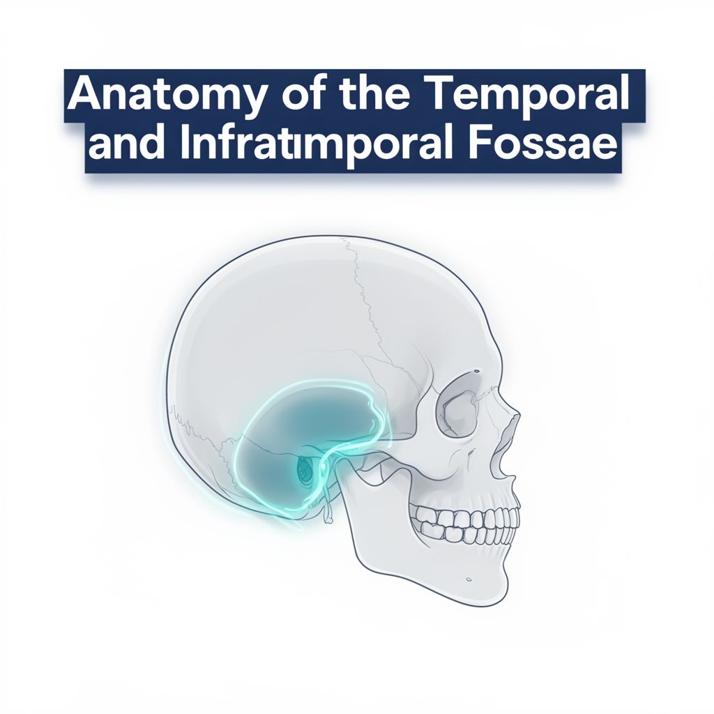
🧠 Anatomy of the Temporal and Infratemporal Fossae

This guide provides a comprehensive overview of the temporal and infratemporal fossae, two crucial anatomical regions of the skull. Understanding these areas is fundamental for studying head and neck anatomy, as they house vital structures and serve as important landmarks for clinical practice.


1. The Temporal Fossa 🕰️

The temporal fossa is a depressed area located on each side of the skull within the temporal region. It is situated between the temporal lines and the zygomatic arch, positioned superiorly to the infratemporal fossa.

1.1. Boundaries of the Temporal Fossa ✅

The temporal fossa is defined by specific bony landmarks:

  • Postero-Superiorly: Superior temporal line
  • Inferiorly: Infratemporal crest
  • Anteriorly: Frontal process of the zygomatic bone
  • Laterally: Zygomatic arch
  • Floor: Formed by the articulation of four bones: frontal, parietal, temporal, and sphenoid.

1.2. The Pterion: A Critical Landmark ⚠️

Within the floor of the temporal fossa lies the pterion.

  • 📚 Definition: The pterion is the thinnest part of the lateral wall of the skull. It is the point where the anteroinferior corner of the parietal bone articulates with the greater wing of the sphenoid bone.
  • Clinical Significance: The pterion is clinically important because it directly overlies the anterior division of the middle meningeal artery and vein. Trauma to this area can lead to severe intracranial hemorrhage, making its precise location critical for medical professionals.

1.3. Contents of the Temporal Fossa 🎯

The primary contents of the temporal fossa include:

  • 1️⃣ Temporalis Muscle:
    • Description: A fan-shaped muscle originating from the superior temporal line. Its fibers converge inferiorly to form a strong tendon.
    • Attachment: The tendon attaches to the anterior surface of the coronoid process of the mandible.
    • Function: A powerful elevator and retractor of the mandible, playing a key role in mastication (chewing).
    • Innervation: Deep temporal nerves (branches of the mandibular nerve, CN V3).
    • Blood Supply: Deep temporal arteries and the superficial temporal artery.
  • 2️⃣ Temporalis Fascia: Covers the temporalis muscle.
  • 3️⃣ Deep Temporal Nerves and Vessels: Supply the temporalis muscle.

1.4. Clinical Significance of the Temporal Fossa 💡

Understanding the temporal fossa is crucial for several reasons:

  • Anatomical Boundaries: Essential for accurate diagnosis and surgical planning.
  • Muscle Function: Knowledge of the temporalis muscle's anatomy, innervation, and blood supply helps in assessing and treating jaw disorders (e.g., temporomandibular joint dysfunction).
  • Pterion: Its location overlying the middle meningeal artery is vital for preventing complications during head trauma or neurosurgical interventions.
  • Connection to Infratemporal Fossa: This continuity is fundamental for grasping the functional anatomy of mastication and understanding the pathways of various neurovascular structures.

2. The Infratemporal Fossa ⬇️

The infratemporal fossa is an irregularly shaped space located on the side of the skull, positioned below the zygomatic arch and deep to the ramus of the mandible. It is situated directly below the temporal fossa and is continuous with it.

2.1. Why Study the Infratemporal Fossa? 📈

This region is critically important due to its complex contents and connections:

  • 1️⃣ Anatomical Link: It links major head and facial regions.
  • 2️⃣ Neurovascular Hub: Contains key neurovascular structures, including the mandibular nerve (CN V3), maxillary artery, and pterygoid plexus of veins. These support blood supply and nerve function throughout the head and face.
  • 3️⃣ Mastication Muscles: Houses essential muscles for chewing (lateral and medial pterygoid muscles).
  • 4️⃣ Clinical & Surgical Relevance: Knowledge of this fossa is critical for various clinical and surgical procedures, especially when working near the middle meningeal artery.
  • 5️⃣ Safe Outcomes: Understanding this area helps ensure safe surgical outcomes and aids in managing jaw function, pain, and vascular issues.

2.2. Boundaries of the Infratemporal Fossa ✅

The infratemporal fossa has the following boundaries:

  • Laterally: Ramus of the mandible
  • Medially: Lateral pterygoid plate
  • Anteriorly: Posterior aspect of the maxilla
  • Posteriorly: Tympanic plate and the mastoid and styloid processes of the temporal bone
  • Superiorly: Greater wing of the sphenoid (forming its roof)
  • Inferiorly: Where the medial pterygoid muscle attaches to the mandible near its angle.

2.3. Communications of the Infratemporal Fossa 🌐

The infratemporal fossa is not isolated but communicates with several other regions:

  • 1️⃣ Temporal Fossa: Through a gap deep to the zygomatic arch.
  • 2️⃣ Cranial Cavity: Via the foramen ovale, foramen spinosum, and foramen lacerum.
  • 3️⃣ Orbit: Through the inferior orbital fissure.
  • 4️⃣ Pterygopalatine Fossa: Through the pterygomaxillary fissure.

2.4. Foramina in the Infratemporal Fossa (Summary) 🕳️

Several foramina open into or from the infratemporal fossa, serving as passages for vital structures:

  • Foramen Spinosum: Passage for the middle meningeal artery into the middle cranial fossa.
  • Foramen Ovale: Passage for the mandibular nerve (CN V3) and accessory meningeal artery.
  • Pterygomaxillary Fissure: A medial cleft leading into the pterygopalatine fossa, containing terminal branches of the maxillary artery.
  • Inferior Orbital Fissure: Leads anteriorly into the orbit. Its contents include the zygomatic branch of the maxillary nerve (CN V2), infraorbital vessels, and the inferior ophthalmic vein (which communicates with the pterygoid plexus).

2.5. Contents of the Infratemporal Fossa 🎯

The infratemporal fossa is rich in structures, including:

  • 1️⃣ Muscles of Mastication:
    • Lower portion of Temporalis Muscle: While most of the temporalis is in the temporal fossa, its lower part extends into the infratemporal fossa.
    • Lateral Pterygoid Muscle: Originates from the lateral pterygoid plate and greater wing of the sphenoid; inserts onto the neck of the mandible and articular disc of the TMJ. Action: Protrudes the mandible.
    • Medial Pterygoid Muscle: Originates from the medial surface of the lateral pterygoid plate and tuberosity of the maxilla; inserts onto the medial surface of the ramus and angle of the mandible. Action: Closes the jaw with bilateral contraction.
  • 2️⃣ Nerves:
    • Mandibular nerve (CN V3) and its branches.
    • Chorda tympani.
    • Otic ganglion.
  • 3️⃣ Vessels:
    • Maxillary artery.
    • Pterygoid plexuses of veins (directly connected to the cavernous sinus and drain the eye and its locality).

This detailed exploration highlights the intricate anatomy and profound clinical importance of the temporal and infratemporal fossae, essential knowledge for any student of human anatomy.

Kendi çalışma materyalini oluştur

PDF, YouTube videosu veya herhangi bir konuyu dakikalar içinde podcast, özet, flash kart ve quiz'e dönüştür. 1.000.000+ kullanıcı tercih ediyor.

Sıradaki Konular

Tümünü keşfet
Skeletal Muscle Structure and Neuromuscular Junction

Skeletal Muscle Structure and Neuromuscular Junction

This audio summary provides an academic overview of skeletal muscle terminology, morphology, filament and sarcomere structure, innervation, and the intricate process of excitation-contraction coupling.

7 dk Özet 18 12
Neuron Types and Nervous System Structures

Neuron Types and Nervous System Structures

This summary explores the diverse morphology and classification of neurons, their fundamental components, synaptic connections, and the crucial role of glial cells and peripheral nervous system structures.

6 dk Özet 25 15
Male Reproductive Anatomy and Gonad Descent

Male Reproductive Anatomy and Gonad Descent

Explore the gross anatomy of the male reproductive system, accessory glands, and the embryological development and descent of gonads, including clinical correlations.

7 dk Özet 15
The Visual Pathway, Reflexes, and Eye Movements

The Visual Pathway, Reflexes, and Eye Movements

An academic overview of the visual pathway from retina to cortex, including visual field processing, magnocellular and parvocellular streams, light and accommodation reflexes, and conjugate eye movements.

8 dk Özet 25 15
The Anatomy and Physiology of the Human Heart

The Anatomy and Physiology of the Human Heart

Explore the intricate structure and function of the human heart, covering its location, chambers, valves, and the electrical conduction system that drives its rhythmic beat.

Özet 25 15
The Vestibular System and Auditory Pathway

The Vestibular System and Auditory Pathway

An academic overview of the vestibular system's role in balance and spatial orientation, and the auditory pathway's function in sound perception, detailing their anatomical structures and neural processing.

8 dk Özet 25 15
Gastrointestinal Tract Blood Flow and Movements

Gastrointestinal Tract Blood Flow and Movements

Explore the intricate details of gastrointestinal blood circulation, including splanchnic flow, anatomical supply, regulatory factors, and the mechanics of propulsive and mixing movements within the GI tract.

9 dk Özet 15
Human Brain Anatomy: Ventricles, Terminology, and Structures

Human Brain Anatomy: Ventricles, Terminology, and Structures

Explore the intricate anatomy of the human brain, covering the ventricular system, essential directional terms, embryonic development, and the major divisions and structures of the adult brain.

25 15