Study Material: The Vestibulocochlear System: Balance and Hearing
Source Information: This study material has been compiled from a lecture audio transcript and copy-pasted text provided by the user.
Introduction to the Vestibulocochlear System 📚
The vestibulocochlear nerve (Cranial Nerve VIII) is a crucial sensory nerve responsible for both balance and hearing. It is composed of axons from bipolar neurons located in the vestibular and spiral ganglia. The inner ear, where these functions originate, consists of a complex system of fluid-filled chambers:
- Bony Labyrinth: Contains perilymph, a fluid similar to extracellular fluid.
- Membranous Labyrinth: Housed within the bony labyrinth, it contains endolymph, a potassium-rich, sodium-poor fluid resembling intracellular fluid.
I. The Vestibular System: Balance and Spatial Orientation 🧭
The vestibular system is responsible for detecting head movements and position, crucial for maintaining balance and spatial orientation.
1. Anatomy of the Vestibular Labyrinth ✅
The vestibular labyrinth includes:
- Utricle and Saccule: Each contains a specialized sensory epithelium called a macula.
- Three Semicircular Ducts: Each duct has an enlargement at its base called an ampulla, which contains a sensory structure called a crista.
- Vestibular Ganglion: Connects these five neuroepithelial end organs (maculae and cristae) to the vestibular nuclei in the brainstem.
2. Static Labyrinth: Head Position and Linear Acceleration 🧍
The maculae of the utricle and saccule are the primary receptors for static head position and linear acceleration.
- Utricular Macula: Primarily horizontal, sensitive to horizontal linear acceleration and head tilts.
- Saccular Macula: Primarily vertical, sensitive to vertical linear acceleration.
- Function: They signal the head's position relative to the trunk, enabling compensatory movements to maintain the center of gravity.
- Hair Cells: These sensory cells have stereocilia and one long kinocilium. They are embedded in a gelatinous matrix containing otoconia (calcium carbonate crystals, or "ear sand").
- Movement of the kinocilium away from the stereocilia facilitates depolarization.
- A striola divides the macula, creating a mirror arrangement of hair cell polarities.
- Vestibulospinal Tracts:
- Lateral Vestibulospinal Tract (Deiters-Lat Vestibular Nuc.): Ipsilateral, located in the anterior funiculus. Primarily activates antigravitational (extensor) muscles, crucial for maintaining posture. Involved in the eye-righting reflex (Deitero-ocular pathway).
- Medial Vestibulospinal Tract: Originates from medial and inferior vestibular nuclei. Functions in the head-righting reflex (HRR).
3. Righting Reflexes 💡
- Head-Righting Reflex (HRR): Maintained by the medial vestibular tract. Keeps the head in a stationary position (to keep eyes focused) relative to body movement (e.g., sideways or forward).
- Eye-Righting Reflex: Works through the Medial Longitudinal Fasciculus (MLF). Provides contralateral torsional movements of the eyeballs to fix an object on the foveola (also known as rotational VOR).
4. Kinetic Labyrinth: Head Movements and Angular Acceleration 🔄
The cristae within the ampullae of the semicircular ducts are sensitive to angular acceleration (rotational head movements).
- Structure: Kinocilia penetrate into a gelatinous mass called the cupula, which is bonded to the opposite wall of the ampulla.
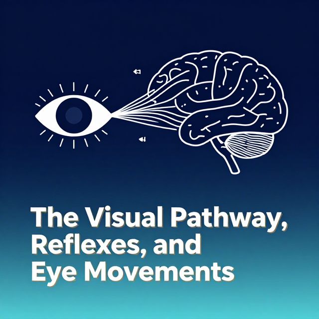
- Function: Connected to the vestibular nuclei and flocconodular lobe, they maintain compensatory eye movements in response to head movements through the Vestibulo-Ocular Reflex (VOR).
5. Vestibulo-Ocular Reflex (VOR) 👀
The VOR ensures that despite head movement, the eyes remain focused on an object by moving in the opposite direction to the head.
- Mechanism: The vestibular system detects head movement and informs the Paramedian Pontine Reticular Formation (PPRF) for horizontal conjugate eye movements and the nucleus of Cajal for vertical conjugate eye movements.
- Voluntary Control: The PPRF is also voluntarily controlled by the contralateral Frontal Eye Field (FEF), not solely by vestibular nuclei.
6. Clinical Applications of VOR 🩺
- Oculocephalic Reflex (Doll's Eyes Reflex): An application of the VOR used for neurological examination.
- Procedure: When the head is rotated, the eyes should deviate contralaterally (opposite to head movement).
- Interpretation: Contralateral eye deviation confirms brainstem integrity. Spontaneous return of eyes to midline confirms cerebrum integrity.
- Caloric Reflex Test: Evaluates the VOR pathway and brainstem function, especially in comatose patients or to assess asymmetrical peripheral vestibular function.
- Procedure: Cold or hot water is irrigated into the ear canal.
- Example (Right Hot Water): Causes eyes to slowly deviate to the left (via VOR), then quickly snap back to the right (cortical stimulation). This slow-then-fast movement is called vestibular nystagmus.
7. Vestibulocortical Connections 🧠
Vestibular signals project from the vestibular nucleus to the contralateral ventral posterior medial (VPM) nucleus of the thalamus, and then to the insula and temporoparietal cortex.
8. Vestibular Dysfunctions ⚠️
- Unilateral Vestibular Disease: (e.g., acoustic neuroma)
- Eyeball torsions towards the disease side (due to unopposed activity of the intact side).
- Head tilt to the disease side.
- Tendency to fall to the disease side (due to lateral vestibulospinal tract insufficiency).
- Vertigo: An illusion or abnormal perception of motion.
- Causes: Any disease affecting the vestibular system, such as otitis media, trauma, Meniere’s disease, acoustic neuroma, cerebellopontine tumors, or cholesteatoma.
- Vestibular Ataxia: Dysfunction of the vestibulospinal tract.
- Features (📊 compared to Cerebellar & Sensory Ataxia):
- Limb Coordination (Finger-to-Nose): Normal
- Gait: Staggers toward the lesion side.
- Romberg Test: Positive (falls with eyes closed).
- Vertigo: Present (often severe).
- Features (📊 compared to Cerebellar & Sensory Ataxia):
- Lateral Medullary Syndrome: Damage to vestibular nuclei can lead to vertigo, often with initial vomiting, and symptoms of unilateral labyrinth disconnection.
II. The Auditory Pathway: Sound Perception 👂
The auditory pathway processes sound waves into neural signals, allowing for sound perception.
1. Anatomy of the Cochlea ✅
- Cochlear Nerve: Formed by axons of bipolar neurons in the spiral ganglion.
- Modiolus: The central bony pillar, axis of the internal acoustic meatus where the cochlear nerve originates.
- Osseous Spiral Canal: Contains three fluid-filled compartments:
- Scala Vestibuli: Contains perilymph.
- Scala Media (Cochlear Duct): Contains endolymph.
- Scala Tympani: Contains perilymph.
- Organ of Corti: Located on the basilar membrane within the scala media.
- Contains inner and outer hair cells and supporting cells.
- Hair cells have stereocilia but no kinocilia.
- Covered by the tectorial membrane.
2. Sound Transduction 🎶
- Tympanic Membrane vibrates.
- Ossicle Chain (malleus, incus, stapes) transmits vibrations.
- Oval Window receives vibrations, transferring energy to perilymph in the scala vestibuli.
- Vibrations cause movement of the basilar membrane.
- Tonotopic Recognition: The basilar membrane is tonotopically organized:
- Base: Shorter membrane, vibrated by high-frequency sounds.
- Apex: Longest membrane, vibrated by low-frequency sounds.
3. Central Auditory Pathways 🧠
Auditory signals travel through a complex series of nuclei and tracts:
- Spiral Ganglion (first-order neurons).
- Cochlear Nucleus:
- Dorsal Nucleus: Processes pitch information.
- Ventral Nucleus: Processes intensity information.
- Trapezoid Body.
- Superior Olivary Nucleus: Contains binaural neurons that integrate intensity and timing of sounds from both ears, crucial for detecting the spatial direction of incoming sounds.
- Lateral Lemniscus: Contains the lateral lemniscus nucleus, involved in reflex arcs (e.g., for motor nuclei of CN V and VII) and the startle response.
- Inferior Colliculus: Integrates all auditory information and contributes to the tectospinal tract.
- Inferior Brachium.
- Medial Geniculate Body (of Thalamus): Gives rise to auditory radiation.
- Primary Auditory Cortex: Located in the superior temporal gyrus (transverse temporal gyri or Heschl's gyrus).
- Tonotopic arrangement is preserved here.
- Removal of this area results in partial deafness and loss of judgment of sound direction and distance.
4. Descending (Efferent) Auditory Pathways ⬇️
These pathways modulate auditory processing:
- Originate from the primary auditory cortex, medial geniculate body, and inferior colliculus.
- Project to the superior olivary nucleus via the olivocochlear bundle.
- Cholinergic efferent fibers target the hair cells of the cochlea, possibly enhancing the detection of faint sounds.
5. Types of Deafness and Lesions 👂
- Conductive Deafness: Problems with sound transmission to the inner ear (e.g., earwax, ossicle damage).
- Sensorineural Deafness: Damage to the inner ear (cochlea) or auditory nerve.
- Presbycusis: The most common form in the elderly, characterized by loss of high-frequency sounds due to deterioration of the Organ of Corti in the basal turn.
- Lesions in the Auditory Pathway:
- Cochlear Nerve Lesion: Results in severe hearing loss.
- Higher Pathway Lesions (e.g., Superior Olivary Nucleus): Can cause loss of intensity and spatial localization of sound.
- Central Pathway Lesions: Due to bilateral projection to both auditory cortices, these typically result in partial but bilateral hearing loss, rather than complete unilateral deafness.