This study material has been compiled from various sources, including a lecture audio transcript and copy-pasted text notes.
🧠 Neuron Types and Nervous System Organization: A Comprehensive Study Guide
🎯 Introduction to Neurons
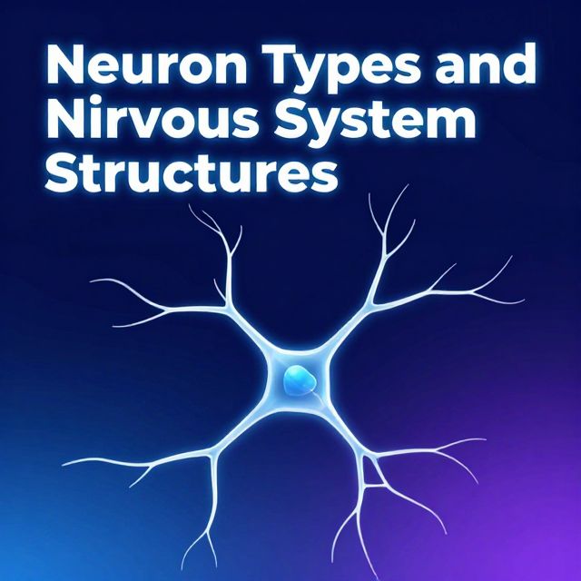
Neurons are the fundamental structural and functional units of the nervous system. They are specialized cells responsible for transmitting electrical and chemical signals throughout the body, enabling communication between different parts of an organism. This intricate communication underlies all nervous system functions, from basic reflexes to complex cognitive processes. Understanding the diverse types of neurons, their specialized structures, synaptic connections, and the crucial support provided by glial cells is essential for comprehending the complexity of neural circuits.
This guide will delve into the primary classifications of neurons based on their morphology, detail key cellular components, explain the various types of synapses, and describe the organization of peripheral nervous system elements, including Schwann cells, myelinated axons, and ganglia.
1️⃣ Neuron Morphology and Classification
Neurons exhibit remarkable diversity in their shapes and sizes, which directly correlates with their specific functions. The classification of neurons is primarily based on the number of processes (extensions) that emanate from the cell body.
📚 Key Term: Perikaryon (Soma) The cell body of a neuron, also known as the soma, is the metabolic center of the neuron. It contains the nucleus and most of the cell's organelles.
Types of Neurons Based on Processes:
-
1. Multipolar Neurons
- ✅ Description: These are the most common type of neurons. They are characterized by having multiple dendrites (tree-like extensions that receive signals) and a single axon (a long projection that transmits signals away from the cell body).
- ✅ Location & Function:
- Motor Neurons: Transmit signals from the central nervous system (CNS) to muscles and glands, initiating movement or secretion.
- Interneurons: Found entirely within the CNS, they connect other neurons, facilitating complex neural circuits.
- Sympathetic Ganglia: Found in the autonomic nervous system, involved in 'fight or flight' responses.
- 💡 Insight: Their extensive dendritic trees allow them to integrate signals from numerous other neurons.
-
2. Pseudounipolar Neurons
- ✅ Description: These neurons have a unique structure where a single process emerges from the cell body and then immediately bifurcates (splits) into two branches. One branch extends towards the periphery (e.g., skin, muscles) to receive sensory input, and the other projects into the CNS.
- ✅ Location & Function:
- Sensory Neurons: Primarily found in sensory ganglia (e.g., dorsal root ganglia), they transmit sensory information (touch, pain, temperature) from the body's periphery to the CNS.
- 💡 Insight: Despite appearing to have one process, they are functionally bipolar, with one part acting as a dendrite (receiving sensory input) and the other as an axon (transmitting it to the CNS).
-
3. Bipolar Neurons
- ✅ Description: These neurons are less common and possess two processes extending from opposite ends of the cell body: one dendrite and one axon.
- ✅ Location & Function:
- Specialized Sensory Organs: Typically found in the retina of the eye, the olfactory epithelium (smell), and the inner ear (hearing and balance), where they transmit specific sensory information.
2️⃣ Key Cellular Components of Neurons
Beyond their overall morphology, neurons contain specialized internal structures crucial for their function.
- 📚 Perikaryon (Soma): As mentioned, this is the cell body, containing the nucleus, which houses the neuron's genetic material, and other organelles vital for cell maintenance and function.
- 📚 Nissl Bodies:
- ✅ Description: These are prominent aggregates of rough endoplasmic reticulum (RER) and free ribosomes found within the perikaryon and dendrites (but generally not in the axon or axon hillock). When stained, they appear as dark granular structures.
- ✅ Function: Neurons are highly active cells that synthesize a large amount of protein (e.g., neurotransmitters, enzymes, structural proteins). Nissl bodies are the primary sites of this high rate of protein synthesis, essential for neuronal growth, repair, and the production of neurotransmitters.
- 📚 Glia (Neuroglia):
- ✅ Description: These are non-neuronal cells that provide essential support, protection, and nourishment to neurons. They are far more numerous than neurons and play critical roles in maintaining the nervous system's environment.
- ✅ Function: Glial cells perform various functions, including forming myelin, regulating the chemical environment, providing metabolic support, and participating in immune responses within the nervous system.
- 📚 Neuropil:
- ✅ Description: This term refers to the dense, intricate network of unmyelinated axons, dendrites, and glial cell processes found in the gray matter of the CNS. It's the region where most synaptic connections occur.
- ✅ Function: The neuropil is the primary site of information processing and communication between neurons in the CNS.
- 📚 Axon:
- ✅ Description: A single, long projection extending from the perikaryon, specialized for transmitting electrical signals (action potentials) away from the cell body to other neurons or effector cells.
- 📚 Axon Terminal:
- ✅ Description: The specialized end portion of an axon, often branching into several terminals.
- ✅ Function: This is the presynaptic part of a synapse, where neurotransmitters are stored in vesicles and released into the synaptic cleft to communicate with the postsynaptic cell.
3️⃣ Synaptic Transmission and Types of Synapses
Neuronal communication primarily occurs at specialized junctions called synapses. These are critical for the flow of information throughout the nervous system.
📚 Key Term: Synapse A synapse is a specialized junction between two neurons (or a neuron and an effector cell like a muscle or gland) where electrical or chemical signals are transmitted.
Types of Synapses Based on Location:
Synapses are classified based on which parts of the neurons are involved in the connection:
- 1. Axodendritic Synapses:
- ✅ Description: The most common type, where the axon terminal of one neuron communicates with a dendrite (or dendritic spine) of another neuron.
- ✅ Function: Typically excitatory, but can also be inhibitory, influencing the postsynaptic neuron's firing.
- 2. Axosomatic Synapses:
- ✅ Description: Occur when the axon terminal of one neuron forms a synapse directly on the cell body (soma) of another neuron.
- ✅ Function: Often inhibitory, having a powerful influence on the postsynaptic neuron's ability to generate an action potential because of its proximity to the axon hillock (the site of action potential initiation).
- 3. Axoaxonic Synapses:
- ✅ Description: Involve the axon terminal of one neuron synapsing with the axon of another neuron, usually near its terminal.
- ✅ Function: These synapses primarily modulate the amount of neurotransmitter released by the postsynaptic axon terminal, providing a mechanism for presynaptic inhibition or facilitation. They don't directly cause the postsynaptic neuron to fire but regulate its output.
4️⃣ Glial Support in the Peripheral Nervous System (PNS)
Glial cells are indispensable for the proper functioning of neurons. In the PNS, specific glial cells play crucial roles.
-
📚 Schwann Cells:
- ✅ Description: These are the principal glial cells of the PNS.
- ✅ Function:
- Myelination: Schwann cells form the myelin sheath around axons in the PNS. Myelin is a fatty, insulating layer that significantly increases the speed of nerve impulse conduction. A single Schwann cell can myelinate only one segment of one axon.
- Support for Unmyelinated Axons: Schwann cells also envelop unmyelinated axons, providing metabolic support and protection, although they do not form a thick myelin sheath.
- 📊 Myelinated Axon Structure:
- Outer Collar: The outermost layer of the Schwann cell, containing its nucleus and most of its cytoplasm.
- Inner Collar: The tightly wrapped layers of Schwann cell membrane that form the myelin sheath directly around the axon.
- Schmidt-Lanterman Clefts: These are small, oblique pockets of Schwann cell cytoplasm within the compact myelin layers. They are thought to facilitate metabolic exchange between the outer and inner parts of the Schwann cell and the axon.
- 💡 Insight: Myelination is crucial for rapid signal transmission, allowing for quick responses and efficient communication over long distances.
-
📚 Satellite Cells:
- ✅ Description: These are small glial cells that surround the cell bodies of neurons in PNS ganglia.
- ✅ Function: They provide structural and metabolic support to the ganglion neurons, helping to regulate the microenvironment around the neuronal cell bodies.
5️⃣ Ganglia and Peripheral Nerve Organization
Ganglia are vital relay stations in the PNS, housing collections of neuron cell bodies.
📚 Key Term: Ganglion (plural: Ganglia) A ganglion is a cluster of neuron cell bodies located outside the central nervous system.
Types of Ganglia:
-
1. Sympathetic Ganglion:
- ✅ Location: Part of the autonomic nervous system, typically found in chains alongside the vertebral column or closer to target organs.
- ✅ Neuron Type: Primarily contains multipolar neurons.
- ✅ Function: Involved in the 'fight or flight' response, mediating involuntary actions like increasing heart rate, dilating pupils, and diverting blood flow.
-
2. Sensory Ganglion (e.g., Dorsal Root Ganglion):
- ✅ Location: Found along the dorsal (posterior) roots of spinal nerves.
- ✅ Neuron Type: Contains the cell bodies of pseudounipolar neurons.
- ✅ Function: Transmit sensory information (e.g., touch, pain, temperature, proprioception) from the periphery to the CNS.
-
📚 Peripheral Nerve:
- ✅ Description: A peripheral nerve is a bundle of many axons (both myelinated and unmyelinated) that are encased in various layers of connective tissue.
- ✅ Organization: Within a nerve, individual axons are surrounded by Schwann cells. These bundles of axons are then grouped together, forming the macroscopic structure of a nerve.
- ✅ Function: Peripheral nerves serve as the communication highways, carrying motor commands from the CNS to muscles and glands, and sensory information from the body back to the CNS.
✅ Conclusion
The nervous system's remarkable ability to process and transmit information relies on the diverse types of neurons and their intricate organization. From the multipolar neurons driving motor functions to the pseudounipolar neurons conveying sensory input, each neuron type is structurally adapted for its specific role. The perikaryon, with its protein-synthesizing Nissl bodies, serves as the metabolic center, while axons and dendrites form the communication pathways. Synapses, whether axodendritic, axosomatic, or axoaxonic, are the precise points of information transfer. Furthermore, the essential support provided by glial cells, such as Schwann cells in myelination and satellite cells in ganglia, underscores the collaborative nature of nervous tissue. This sophisticated architecture enables the nervous system to perform its complex functions, from basic reflexes to higher cognitive processes.