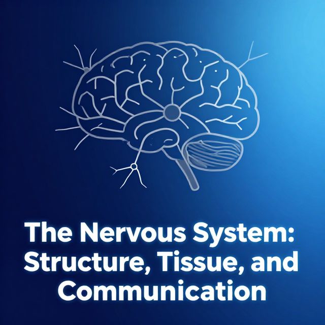
🧠 The Nervous System and Nervous Tissue: A Comprehensive Study Guide
This study material has been compiled and organized from a lecture audio transcript and copy-pasted text sources to provide a clear and structured overview of the nervous system.
📚 Introduction to the Nervous System
The nervous system is an incredibly complex biological network responsible for controlling and coordinating all bodily functions. It enables us to perceive our environment, process information, and generate appropriate responses. Understanding its fundamental structure, cellular components, and the mechanisms of signal transmission is crucial for grasping its physiological roles.
1️⃣ Basic Structure and Functional Divisions
The nervous system is organized into distinct anatomical and functional components.
1.1 Anatomical Divisions
✅ Central Nervous System (CNS): * Comprises the brain and spinal cord. * Acts as the main processing center for all information. ✅ Peripheral Nervous System (PNS): * Includes all neural structures outside the brain and spinal cord. * Connects the CNS to the rest of the body, including organs, limbs, and skin.
1.2 Functional Divisions
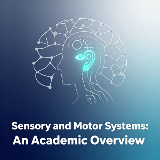
The nervous system performs three primary functions:
- Sensation: Detecting internal and external stimuli (e.g., touch, temperature, light).
- Integration: Processing and interpreting sensory information to make decisions.
- Generating Response: Executing appropriate actions based on integrated information (e.g., muscle contraction, gland secretion).
1.3 Gray and White Matter
Nervous tissue is further categorized by its appearance and cellular composition:
- Gray Matter:
- Contains neuronal cell bodies, dendrites, unmyelinated axons, and glial cells.
- Forms nuclei in the CNS and ganglia in the PNS.
- Primarily involved in processing information.
- White Matter:
- Composed mainly of myelinated axons.
- Forms tracts in the CNS and nerves in the PNS.
- Primarily responsible for transmitting signals rapidly between different parts of the brain and body.
2️⃣ Nervous Tissue: Neurons, Glia, and Myelination
Nervous tissue consists of two major cell types that work together to facilitate communication.
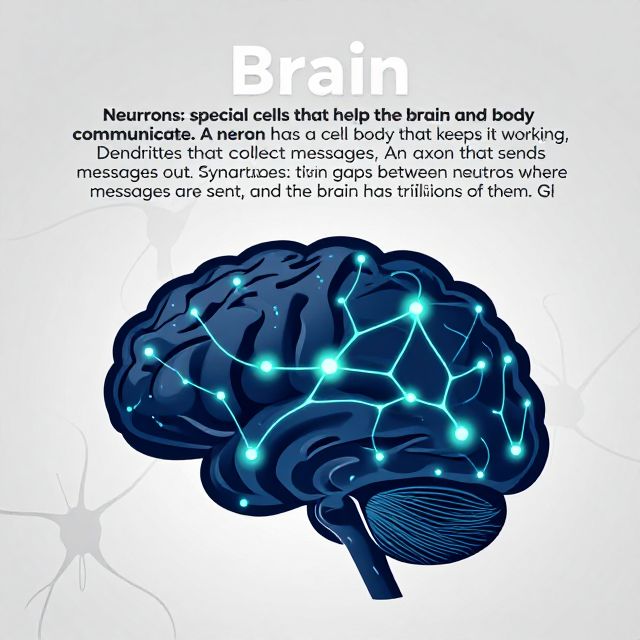
2.1 Neurons
📚 Neurons: The fundamental units of the nervous system responsible for communication through electrical signals. They transmit information throughout the body.
2.2 Glial Cells (Neuroglia)
📚 Glial Cells: Supporting cells that maintain the environment around neurons. They provide structural support, insulation, and metabolic assistance.
- Functions:
- Support and protect neurons.
- Maintain homeostasis.
- Form myelin.
- Participate in neural activity.
2.3 Myelination
Myelination is a critical process where a fatty insulating sheath, called myelin, surrounds axons. This significantly increases the speed of electrical signal conduction.
- Myelin Formation:
- In the PNS: Formed by Schwann cells. Each Schwann cell typically myelinates a single axon.
- In the CNS: Formed by oligodendrocytes. A single oligodendrocyte can myelinate segments of multiple axons simultaneously.
- Process: Glial cells form myelin by extending processes that rotate around an axon over 100 times, creating a multi-layered sheath.
- Importance: Essential for efficient and rapid nerve impulse transmission.
3️⃣ Neuronal Communication: Resting Potential and Action Potential
Neurons communicate through electrical signals generated by changes in their membrane potential.
3.1 Resting Membrane Potential (RMP)
📚 Resting Membrane Potential (RMP): The electrical potential difference across the neuronal membrane when the neuron is not actively signaling. It is typically negative inside the cell relative to the outside (e.g., -70 mV).
- Maintenance: Maintained by the differential distribution of ions (primarily Na⁺, K⁺, Cl⁻, and large organic anions) and the activity of the Na⁺/K⁺ pump.
- Clinical Relevance: Disruptions to RMP can have significant physiological consequences:
- ⚠️ Severe Hyponatremia:
- ↓ Plasma Na⁺ → ↓ osmolarity → brain edema → impaired neuronal function → confusion/coma.
- ⚠️ Hypokalemia:
- ↓ Extracellular K⁺ → hyperpolarization (membrane becomes more negative) → unstable excitability → abnormal firing → muscle cramps.
- ⚠️ Dehydration:
- Fluid loss → electrolyte imbalance → disrupted RMP → unstable signaling.
- ⚠️ Severe Hyponatremia:
3.2 The Action Potential (AP)
📚 Action Potential (AP): A brief, rapid, and reversible change in the membrane potential that constitutes a signal. It is the basis of rapid neuronal communication.
- Mechanism: Involves a rapid depolarization (becoming positive inside) followed by repolarization (returning to negative) and a brief hyperpolarization.
- Measurement: A potential is a distribution of charge across the cell membrane, measured in millivolts (mV). The standard compares the inside of the cell relative to the outside (outside is considered zero).
- Factors Affecting AP Generation and Propagation:
- ⚠️ ATP Depletion:
- ↓ ATP → Na⁺/K⁺ pump failure (pump requires ATP) → loss of ion gradients → membrane depolarization → no AP generation.
- ⚠️ Hypoxia:
- ↓ O₂ → ↓ ATP production → pump failure → neuronal depolarization → dysfunction.
- ⚠️ Glucose Deficiency:
- ↓ Glucose → ↓ ATP → pump dysfunction → loss of RMP → neuronal damage.
- 💡 Local Anesthetics:
- Block Na⁺ channels → prevent depolarization → no AP propagation → no pain transmission.
- 💡 Hypercalcemia:
- ↑ Ca²⁺ → stabilized Na⁺ channels → ↑ threshold for AP → ↓ neuronal firing (reduced excitability).
- ⚠️ Vitamin B12 Deficiency:
- ↓ B12 → impaired myelin synthesis → slowed conduction → sensory/motor deficits (neuropathy).
- ⚠️ Multiple Sclerosis (MS):
- Myelin loss → ↓ insulation → impaired saltatory conduction (AP "jumps" between nodes) → slow transmission.
- ⚠️ Chronic Malnutrition:
- Nutrient deficiency → ↓ neurotransmission & myelin integrity → impaired synaptic efficiency → cognitive dysfunction.
- ⚠️ ATP Depletion:
4️⃣ Communication Between Neurons: Synaptic Transmission
For a neuron to generate an action potential, it typically receives input from another source, either another neuron or a sensory stimulus.
4.1 Graded Potentials
- Input (from another neuron or sensory stimulus) results in the opening of ion channels in the receiving neuron.
- This leads to a graded potential, which is a localized change in membrane potential.
- The magnitude of the graded potential is directly proportional to the strength of the stimulus.
- If sufficiently strong, graded potentials can summate to reach the threshold for generating an action potential.
4.2 Synapses
📚 Synapses: Specialized junctions where communication between neurons occurs.
- Presynaptic Neuron: The neuron sending the signal.
- Postsynaptic Neuron: The neuron receiving the signal.
- Neurotransmitters: Chemical messengers released from the presynaptic neuron into the synaptic cleft.
- Mechanism: Neurotransmitters bind to receptors on the postsynaptic neuron, altering its membrane potential and either exciting or inhibiting it.
4.3 Clinical Example: Magnesium Deficiency
- ⚠️ Magnesium Deficiency:
- ↓ Mg²⁺ → ↑ Ca²⁺ influx into neurons → ↑ neurotransmitter release → increased neuromuscular irritability (e.g., muscle spasms, tremors).
- This intricate process of synaptic transmission ensures precise and regulated information flow throughout the nervous system.
5️⃣ Conclusion
The nervous system is a highly organized and complex network, divided anatomically into central and peripheral components and functionally into sensation, integration, and response. Its fundamental units, neurons and glial cells, work in concert, with myelination playing a crucial role in signal speed. Neuronal communication relies on the precise regulation of resting membrane potential and the generation of action potentials, which are influenced by various physiological and pathological conditions. Finally, communication between neurons occurs via synaptic transmission, where graded potentials and neurotransmitter release facilitate information transfer. These mechanisms collectively underpin the nervous system's capacity to control and coordinate all bodily functions.