Flash Kartlar
25 kartKarta tıklayarak çevir. ← → ile gez, ⎵ ile çevir.
Tüm kartları metin olarak gör
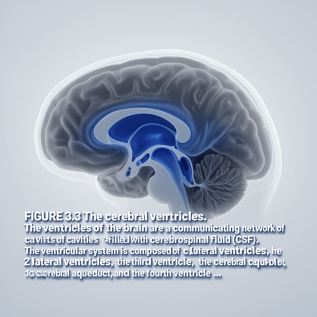
1. What are cerebral ventricles?
Cerebral ventricles are a communicating network of cavities located deep within the brain. These cavities are filled with cerebrospinal fluid (CSF). They are an essential part of the brain's internal structure, facilitating the circulation of CSF.
2. What fluid fills the cerebral ventricles, and what is its primary function?
The cerebral ventricles are filled with cerebrospinal fluid (CSF). This clear fluid surrounds and protects both the brain and the spinal cord. Its primary function is to provide cushioning, nutrient delivery, and waste removal for the central nervous system.
3. List the main components of the ventricular system.
The ventricular system is composed of several key parts. These include two lateral ventricles, the third ventricle, the cerebral aqueduct, and the fourth ventricle. This network ensures a continuous flow of CSF throughout the brain.
4. What is the crucial role of the cerebral aqueduct within the ventricular system?
The cerebral aqueduct plays a crucial role by connecting the third ventricle to the fourth ventricle. This connection is vital for ensuring a continuous and uninterrupted flow of cerebrospinal fluid (CSF) throughout the entire ventricular system. Disruptions here can have significant consequences.
5. Define hydrocephalus and explain its underlying cause.
Hydrocephalus is a medical condition characterized by the accumulation of excess cerebrospinal fluid (CSF) within the brain's ventricular cavities. This buildup occurs when there's an imbalance between CSF production and absorption or a blockage in its flow. The excess fluid increases the size of the ventricles.
6. What are the potential consequences of hydrocephalus if left untreated?
If left untreated, hydrocephalus can lead to serious neurological impairments. The accumulation of excess CSF increases pressure on the brain tissue. This elevated pressure can damage brain cells and interfere with normal brain function, potentially causing developmental delays, cognitive issues, and motor problems.
7. Explain the anatomical directional terms 'anterior' and 'posterior'.
'Anterior' refers to the front or toward the front of the body or a structure. Conversely, 'posterior' means behind or toward the back of the body or a structure. These terms are fundamental for precisely describing locations in anatomy.
8. Differentiate between 'distal' and 'proximal' in anatomical terms.
'Distal' indicates a position away from or farther from the origin or point of attachment of a body part. In contrast, 'proximal' means near or closer to the origin or point of attachment. These terms are often used when describing limbs or appendages.
9. Define 'dorsal' and 'ventral' in general anatomical context.
'Dorsal' refers to the back or upper surface of an organism or structure. For example, the back of the hand is dorsal. 'Ventral' refers to the front or lower surface, such as the belly side of an animal. These terms help describe orientation relative to the main body axis.
10. How are 'rostral' and 'caudal' used specifically in the context of the brain?
In the context of the brain, 'rostral' means toward the front or the nose end. 'Caudal' means toward the back or the tail end. These terms are particularly useful for describing the anterior-posterior axis of the brain, which has a curved orientation.
11. Explain the difference between 'superior' and 'inferior'.
'Superior' denotes a position above or over another structure. For instance, the head is superior to the neck. 'Inferior' signifies a position below or under another structure, such as the feet being inferior to the knees. These terms establish vertical relationships.
12. What do 'lateral' and 'medial' mean in anatomical descriptions?
'Lateral' means toward the side, away from the midline of the body or a structure. For example, the ears are lateral to the nose. 'Medial' means toward the midline or middle, away from the side. The heart is medial to the lungs. These terms describe horizontal positioning.
13. Differentiate between 'bilateral' and 'unilateral' involvement.
'Bilateral' describes something that involves both sides of the body or a structure. For example, bilateral knee pain affects both knees. 'Unilateral' describes involvement of only one side of the body or a structure. A unilateral headache affects only one side of the head.
14. Explain the terms 'ipsilateral' and 'contralateral'.
'Ipsilateral' refers to structures or events occurring on the same side of the body. For instance, an injury affecting the left arm and left leg is ipsilateral. 'Contralateral' refers to structures or events occurring on opposite sides of the body. The right brain hemisphere controls the contralateral, or left, side of the body.
15. What do 'parietal' and 'visceral' refer to in anatomy?
'Parietal' relates to the wall of a body cavity, such as the parietal pleura lining the thoracic cavity. 'Visceral' pertains to the organs located within body cavities, like the visceral pleura covering the lungs. These terms distinguish between the lining and the organ itself.
16. Define 'axial' and 'intermediate' in anatomical terminology.
'Axial' describes something situated around a central axis or the main part of the body, like the axial skeleton. 'Intermediate' means positioned between two other structures or locations. For example, the navel is intermediate to the sternum and the pubic bone.
17. What are the three primary germ layers formed during early embryonic development?
Approximately sixteen days after fertilization, the developing embryo forms three primary germ layers. These fundamental layers are the endoderm, the mesoderm, and the ectoderm. Each layer is destined to give rise to specific tissues and organs in the developing organism.
18. What tissues and structures develop from the endoderm?
The endoderm is the innermost of the three primary germ layers. It primarily forms the lining tissues within the body. This includes the mucosae of the digestive and respiratory systems, as well as glands associated with these systems, such as the liver and pancreas.
19. What does the mesoderm develop into?
The mesoderm is the middle primary germ layer. It develops into a wide variety of tissues and structures throughout the body. Key derivatives include muscle tissue, connective tissues like bone and cartilage, the circulatory system, and the urogenital system.
20. What does the ectoderm give rise to, particularly relevant to the brain?
The ectoderm is the outermost primary germ layer. It gives rise to the skin and its appendages, such as hair and nails. Crucially for our discussion, the ectoderm is also responsible for forming the nervous tissue, including the brain and spinal cord, as well as sensory organs.
21. What are the three broad categories of the adult human brain?
The adult human brain can be broadly categorized into three major divisions. These are the forebrain, the midbrain, and the hindbrain. Each of these categories contains numerous specialized structures responsible for different functions, from basic survival to complex thought.
22. What are the two main divisions of the forebrain?
The forebrain, which is the largest and most complex part of the brain, consists of two main divisions. These are the telencephalon and the diencephalon. Both play critical roles in higher-level cognitive functions, sensory processing, and regulation of bodily states.
23. List key components of the telencephalon and their primary functions.
The telencephalon includes several crucial components. The cerebral cortex is responsible for higher-level functions like thought and language. The basal ganglia are involved in motor control. The hippocampus is vital for memory formation, and the amygdala is central to processing emotions.
24. What structures comprise the diencephalon and what are their primary roles?
The diencephalon consists of the thalamus and the hypothalamus. The thalamus acts as the brain's relay station for sensory information, directing it to appropriate cortical areas. The hypothalamus plays a critical role in regulating essential bodily functions such as hunger, thirst, and temperature, maintaining homeostasis.
25. Describe the main components of the midbrain (mesencephalon) and their functions.
The midbrain, or mesencephalon, comprises the tectum and the tegmentum. The tectum contains the superior and inferior colliculi, involved in visual and auditory reflexes, respectively. The tegmentum includes structures like the periaqueductal gray, substantia nigra, and red nucleus, which are important for motor control and pain modulation.
Bilgini Test Et
15 soruÇoktan seçmeli sorularla öğrendiklerini ölç. Cevap + açıklama.
What fluid fills the cerebral ventricles and surrounds the brain and spinal cord?