This study material has been compiled from a lecture audio transcript and copy-pasted text provided by the user.
❤️ The Human Heart: Anatomy, Function, and Conduction System
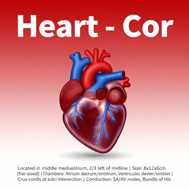
The heart (Latin: Cor) is a vital muscular organ responsible for pumping blood throughout the body. This guide provides a comprehensive overview of its structure, function, and electrical system.
1. General Anatomy and Location 🌍
The heart is strategically located in the middle mediastinum, a central compartment of the thoracic cavity.
- Position: Approximately two-thirds of its mass lies to the left of the body's midline.
- Size: Roughly the size of a clenched fist, measuring about 8 cm (width) x 12 cm (length) x 6 cm (anterior-posterior).
- Weight:
- Males: ~300 g (0.45% of body weight)
- Females: ~250 g (0.40% of body weight)
- Shape: Conical.
- Orientation:
- Base: Faces right, upwards, and backwards.
- Apex: Points forwards, left, and downwards.
Heart Surfaces and Borders 🗺️
The heart presents distinct surfaces and borders:
- Surfaces:
- Facies sternocostalis (Anterior): Primarily formed by the right ventricle.
- Facies diaphragmatica (Inferior): Formed by the posterior walls of both right and left ventricles (clinically known as the "posterior wall").
- Facies pulmonalis (Left): The left surface.
- Facies vertebralis: The posterior part, where the bases of both atria rest against the spine.
- Borders:
- Right Border: Mainly formed by the right atrium.
- Left Border (Obtuse Margin): Mostly formed by the left ventricle, partially by the left auricle.
- Inferior Border (Acute Margin): Mainly formed by the right ventricle, with a small part of the left ventricle.
- Superior Border: Formed by the right and left atria, largely hidden by the ascending aorta and pulmonary trunk.
2. Cardiac Chambers and Internal Structures 🫀
The heart is divided into four main compartments or chambers: two atria (receiving chambers) and two ventricles (pumping chambers).
A. Right Atrium (Atrium Dextrum)
- Structures:
- Sinus Venarum Cavarum: Smooth-walled posterior part where systemic veins (SVC, IVC, coronary sinus) open. Lacks pectinate muscles.
- Tuberculum Intervenosum (of Lower): Small projection on the posterior wall, directs blood from SVC towards the atrioventricular opening.
- Crista Terminalis: Internal ridge separating the smooth sinus venarum cavarum from the rough anterior atrial part. Corresponds externally to the Sulcus Terminalis.
- Mm. Pectinati (Pectinate Muscles): Rough, muscular ridges in the anterior part of the atrium and auricle.
- Auricula Dextra (Right Auricle): An ear-like appendage, lies at the root of the ascending aorta. Increases atrial capacity and is a common site for blood clot formation.
- Fossa Ovalis: A depression on the interatrial septum, remnant of the fetal foramen ovale. Its raised margin is the Limbus Fossae Ovalis.
- Orifices:
- Ostium Venae Cavae Superioris (SVC)
- Ostium Venae Cavae Inferioris (IVC): May have a rudimentary valve (Valvula Eustachii).
- Ostium Sinus Coronarii (Coronary Sinus): May have a rudimentary valve (Valvula Thebesii).
- Ostia Venarum Minimarum (Thebesian veins)
- Ostia Venae Ventriculi Dextri Anteriorum (Anterior cardiac veins)
B. Right Ventricle (Ventriculus Dexter)
- Structures:
- Trabeculae Carneae: Irregular muscular ridges on the inner surface.
- Trabecula Septomarginalis (Moderator Band): A muscular band extending from the interventricular septum to the anterior papillary muscle, part of the heart's conduction system.
- Mm. Papillares (Papillary Muscles): Anterior, posterior, and septal. They attach to the cusps of the tricuspid valve via Chordae Tendineae.
- Crista Supraventricularis: A muscular crest that divides the right ventricular cavity into an inflow portion (pars trabecularis) and an outflow portion (pars glabra).
- Infundibulum / Conus Arteriosus: The smooth-walled outflow tract leading to the pulmonary trunk.
- Orifices:
- Ostium Atrioventriculare Dextrum: Guarded by the Valva Tricuspidalis (Tricuspid Valve), with anterior, posterior, and septal cusps.
- Ostium Trunci Pulmonalis: Guarded by the Valva Trunci Pulmonalis (Pulmonary Valve), a semilunar valve with anterior, right, and left semilunar cusps.
C. Left Atrium (Atrium Sinistrum)
- Characteristics: Smaller than the right atrium, with a thinner muscular wall (low-pressure system).
- Structures:
- Mm. Pectinati: Primarily found in the left auricle.
- Auricula Sinistra (Left Auricle): Lies at the root of the pulmonary trunk. Also a common site for clot formation.
- Valvula Foraminis Ovalis: An occasional narrow tissue fold on the interatrial septum, marking the site of fusion between embryonic septa.
- Orifices:
- Ostia Venarum Pulmonalium: Usually four pulmonary veins terminate here.
D. Left Ventricle (Ventriculus Sinister)
- Characteristics: Has the thickest muscular wall (8-12 mm), approximately three times thicker than the right ventricle, reflecting its role in generating high pressure for systemic circulation.
- Structures:
- Trabeculae Carneae: Prominent muscular ridges.
- Mm. Papillares (Papillary Muscles): Anterior and posterior. Attach to the mitral valve cusps via Chordae Tendineae.
- Aortic Vestibule (Vestibulum Aortae): The smooth-walled outflow tract immediately below the aortic orifice, possessing fibrous walls.
- Orifices:
- Ostium Atrioventriculare Sinistrum: Guarded by the Valva Bicuspidalis / Mitralis (Mitral Valve), with anterior and posterior cusps.
- Ostium Aortae: Guarded by the Valva Aortae (Aortic Valve), a semilunar valve with right, left, and posterior semilunar cusps.
Septa and Sulci 🚧
- Septum Interatriale: Separates the right and left atria.
- Septum Interventriculare: Separates the right and left ventricles, composed of a pars membranacea (connective tissue) and a larger pars muscularis.
- Sulcus Atrioventricularis (Coronary Sulcus): A groove separating the atria from the ventricles, housing the coronary arteries and veins.
- Sulcus Interventricularis (Anterior/Posterior): Grooves marking the external position of the interventricular septum.
3. Crux Cordis (Cross of the Heart) ➕
The Crux Cordis is a significant anatomical landmark located on the lower back side of the heart (diaphragmatic surface).
- Formation: It is the area where the coronary sulcus, the posterior interventricular sulcus, and the interatrial groove meet.
- Clinical Importance:
- The atrioventricular nodal artery, a vital vessel, passes in close proximity to this area.
- It serves as an anastomotic point for the right and left coronary arteries.
4. Fetal vs. Postnatal Circulation 👶➡️🧑
The fossa ovalis in the right atrium is a key remnant of fetal circulation.
A. Fetal Circulation (Prenatal) 🤰
- Oxygenated blood from the placenta enters the fetus via the umbilical vein.
- About half bypasses the liver via the ductus venosus to the inferior vena cava.
- Blood entering the right atrium from the inferior vena cava largely bypasses the non-functioning fetal lungs by flowing through the foramen ovale (a right-to-left shunt) directly into the left atrium.
- Blood from the superior vena cava enters the right ventricle and pulmonary trunk, but most bypasses the lungs by flowing through the ductus arteriosus (another right-to-left shunt) into the aorta.
- Partially oxygenated blood returns to the placenta via the umbilical arteries.
B. Postnatal Circulation (After Birth) 🤱
- With the first breath, pulmonary blood pressure falls, and blood flows into the pulmonary arteries.
- The foramen ovale closes (forming the fossa ovalis) and the ductus arteriosus closes (forming the ligamentum arteriosum), eliminating the fetal shunts and separating pulmonary and systemic circulations.
- The umbilical arteries, vein, and ductus venosus occlude, forming ligaments (e.g., round ligament of the liver, ligamentum venosum).
- Blood now passes through the liver for metabolism.
Clinical Relevance of Fossa Ovalis ⚠️
- Patent Foramen Ovale (PFO): If the foramen ovale does not fully close (found in ~20-25% of adults), it can be associated with paradoxical emboli or cryptogenic stroke.
5. Cardiac Skeleton and Valves 🛡️
A. Cardiac Skeleton
- Composition: Dense fibrous connective tissue.
- Function:
- Provides attachment for the myocardium and all heart valves.
- Electrically separates the atrial myocardium from the ventricular myocardium, ensuring coordinated but distinct contractions.
- The only electrical connection is the Bundle of His, which passes through the Trigonum Fibrosum Dextrum.
- Components:
- Anuli Fibrosi: Four fibrous rings around the heart valves (right, left, aortic, pulmonary).
- Trigonum Fibrosum Dextrum/Sinistrum: Fibrous triangles between the rings.
- Tendo Infundibuli: Connects the right fibrous trigone to the aortic and pulmonary rings.
- Tendo Valvulae V. Cavae Inf. (Tendon of Todaro): A fibrous tendon in the right atrium, part of the boundaries of Koch's Triangle.
B. Cardiac Valves 🚪
Heart valves ensure one-way blood flow.
- Characteristics: Not innervated, avascular, operate based on pressure gradients. Histologically, they are duplications of the endocardium.
- Types:
- Atrioventricular (AV) Valves:
- Valva Tricuspidalis (Tricuspid Valve): Between right atrium and right ventricle. Has three cusps.
- Valva Bicuspidalis / Mitralis (Mitral Valve): Between left atrium and left ventricle. Has two cusps.
- Mechanism: Cusps are attached to papillary muscles via chordae tendineae. During ventricular contraction (systole), papillary muscles contract, tightening the chordae to prevent the cusps from prolapsing into the atria, thus preventing regurgitation.
- Papillary Muscles: Three for the tricuspid valve (anterior, posterior, septal) and two for the mitral valve (anterior, posterior).
- Semilunar Valves:
- Valva Trunci Pulmonalis (Pulmonary Valve): Between right ventricle and pulmonary trunk.
- Valva Aortae (Aortic Valve): Between left ventricle and aorta.
- Mechanism: Each consists of three half-moon-shaped folds (valvulae semilunares). They allow blood to flow out of the ventricles into the great arteries but prevent backflow.
- Structure: Each cusp has a Velum (fibrous lamina), a thinned edge called the Lunula, and a central thickening called the Nodulus Valvulae Semilunaris (of Aranzii), which ensures tight closure.
- Sinus Aortae (of Valsalva): Pockets behind the aortic valve cusps, where the coronary arteries originate.
- Atrioventricular (AV) Valves:
6. Cardiac Cycle: Systole and Diastole ⏱️
The heart's action alternates between relaxation and contraction.
- Diastole (Relaxation):
- The heart relaxes, and the atria and ventricles fill with blood.
- The atria contract (atrial systole) to further fill the ventricles.
- Valves: Semilunar valves are closed; atrioventricular valves are open.
- Systole (Contraction):
- Ventricular Systole: The ventricles contract, ejecting blood into the pulmonary artery and aorta.
- Valves: Atrioventricular valves close (producing the "lub" sound); semilunar valves open.
- Heart Sounds: The characteristic "lub-dub" sound of the heart is caused by the alternating closure of the valves.
7. Heart Wall Layers and Myocardium 💪
The heart wall comprises three layers:
- Endocardium: The inner, hydrophilic surface lining the heart chambers, covering valves and chordae tendineae.
- Myocardium: The thick, muscular middle layer responsible for the heart's pumping action. It consists of striated muscle cells.
- Epicardium: The outermost layer, also known as the visceral layer of the serous pericardium.
Myocardium Types and Thickness 📊
The myocardium is divided into:
- Working Myocardium: Responsible for the contraction of the heart chambers.
- Conductive Myocardium: Forms the heart's electrical conduction system.
Myocardial Thickness:
- Right Atrium: ~2 mm
- Left Atrium: ~3 mm
- Right Ventricle: ~3-5 mm
- Left Ventricle: ~8-12 mm (approximately three times thicker than the right ventricle, due to its role in generating high systemic pressure).
8. Cardiac Conduction System (Systema Conducens Cordis) ⚡
This specialized system of myocardial cells generates and conducts automatic, rhythmic electrical impulses, initiating and coordinating heart contractions.
-
Sinoatrial (SA) Node (Nodus Sinuatrialis / Keith-Flack Node):
- Primary Pacemaker: Generates impulses at ~70 beats/min (sinus rhythm).
- Location: Transversely located under the epicardial wall on the posterior side of the right atrium, near the SVC orifice and superior part of the crista terminalis.
- Blood Supply: In 60% of cases, supplied by branches of the right coronary artery (RCA).
-
Internodal and Interatrial Connections: Pathways that conduct impulses from the SA node to the AV node and across the atria.
-
Atrioventricular (AV) Node (Nodus Atrioventricularis / Aschoff-Tawara Node):
- Secondary Pacemaker: Can generate impulses at ~40-60 beats/min if the SA node fails.
- Location: Sagittally located under the endocardium in the interatrial septum, about 1 cm anterior to the coronary sinus ostium, above the septal cusp of the tricuspid valve.
- Triangle of Koch: The AV node is located within this anatomical triangle, defined by the base of the septal cusp of the tricuspid valve (inferior), the Tendon of Todaro (superior), and the ostium of the coronary sinus (posterior).
- Blood Supply: In 90% of cases, supplied by branches of the RCA.
-
Bundle of His (Fasciculus Atrioventricularis):
- Connection: The only electrical connection between the atria and ventricles.
- Pathway: Originates from the AV node, passes through the fibrous skeleton (Trigonum Fibrosum Dextrum) into the interventricular septum.
-
Right and Left Bundle Branches (Crus Dextrum / Sinistrum):
- The Bundle of His divides into these two branches.
- Right Bundle Branch: Leads excitation to the myocardium of the right ventricle, partly via the Trabecula Septomarginalis (Moderator Band).
- Left Bundle Branch: Leads excitation to the interventricular septum and left ventricular myocardium, dividing into three main fascicles.
-
Purkinje Fibers (Rami Subendocardiales):
- Distribution: Distribute excitation rapidly to the working myocardium of the ventricles.
- Contraction Pattern: Stimulate ventricular walls in a retrograde direction (from apex towards atria), causing the apex to contract first. Papillary muscles are stimulated directly by bundle branches, contracting before ventricular walls to ensure AV valve closure during systole.
- Automaticity: Can generate impulses at ~20-40 beats/min if higher pacemakers fail.
9. Coronary Arteries (Brief Overview) 🩸
The heart receives its own blood supply via the coronary arteries.
- Right Coronary Artery (RCA): Originates from the right aortic sinus of the ascending aorta.
- Branches: Conus branch, Right atrial branch, Right sinoatrial nodal branch, Right ventricular artery, Right marginal artery (RM), Posterior interventricular branch (Posterior Descending Artery - PDA), Branch to AV node, Right atrioventricular branch, Right posterolateral artery (can supply part of the left ventricle).