This study material has been compiled from various sources, including copy-pasted text and a lecture audio transcript, to provide a comprehensive overview of the male reproductive system and gonad development.
📚 Male Reproductive System Anatomy & Gonad Development
🎯 Learning Outcomes
By the end of this topic, you should be able to:
- Describe the gross anatomy of the scrotum, testis, epididymis, ductus deferens, prostate gland, and seminal vesicles.
- Apply gross anatomical, embryological, and microanatomical knowledge of the endocrine and reproductive systems to various clinical correlations.
- Describe the development of the indifferent gonad to form the testis and ovary and their subsequent descent into the scrotum/pelvis.
1. 🔬 Gross Anatomy of the Male Reproductive System
1.1. Testis
The testis is the primary male reproductive organ, responsible for spermatogenesis (sperm production).
- Capsule: Encased by the 📚 tunica albuginea, a dense fibrous capsule.
- It thickens posteriorly to form the 📚 mediastinum testis, which acts as a hilum.
- Internal Structure:
- Divided by septa into approximately 300-400 lobules.
- Each lobule contains highly coiled 📚 seminiferous tubules (~600-1200 total), the sites of sperm production.
- These tubules straighten at the mediastinum testis, forming the 📚 rete testis.
- The rete testis then drains into about 12-14 📚 efferent ductules.
1.2. Scrotum
The scrotum is a sac-like structure that houses the testes, providing a cooler environment essential for spermatogenesis. It consists of several layers, from superficial to deep:
- Skin
- Superficial (dartos) fascia
- External spermatic fascia
- Cremaster muscle and fascia
- Internal spermatic fascia
- Tunica vaginalis (visceral and parietal layers, with a potential space between them)
1.3. Epididymis
Located on the posterolateral aspect of the testis, the epididymis is not suspended by the tunica vaginalis.
- Parts: Consists of a head, body, and tail.
- Function: Crucial for the storage and maturation of sperm, where sperm acquire motility and fertilization ability.
1.4. Ductus (Vas) Deferens
This is an uncoiled continuation of the tail of the epididymis. Its path is as follows:
- Ascends within the spermatic cord.
- Passes through the deep inguinal ring.
- Crosses over the external iliac vessels.
- Courses onto the base of the bladder, where it widens to form the 📚 ampulla.
1.5. Accessory Glands
These glands produce seminal fluid, which mixes with sperm to form semen.
1.5.1. Seminal Vesicles
- Location: Paired glands, posterior to the bladder.
- Contribution: Produce approximately 70% of seminal fluid, rich in fructose (for sperm nutrition), enzymes, and proteins.
- Structure: Slightly coiled lumen with diverticula.
- Ducts: The duct of each seminal vesicle joins the ampulla of the vas deferens to form an 📚 ejaculatory duct.
1.5.2. Prostate Gland
- Location: Unpaired accessory gland, inferior to the bladder, pierced by the prostatic urethra.
- Zones: Divided into four zones:
- Anterior fibromuscular (non-glandular) zone
- Central zone
- 📚 Transitional zone: Typical location for benign prostatic hyperplasia (BPH).
- 📚 Peripheral zone: Typical location for prostate cancers.
- Contribution: Produces about 20% of seminal fluid, which is alkaline to neutralize the acidity of the vagina, aiding sperm survival.
1.5.3. Bulbourethral Glands (Cowper's Glands)
- Location: Paired accessory glands situated in the deep perineal pouch.
- Opening: Open into the penile urethra.
1.6. Penis
The external male organ of copulation.
- Parts: Consists of a root (in the perineum), body, and glans penis.
- Structure: Contains erectile tissues and neurovasculature, covered by skin.
- Features: May include a 📚 prepuce (foreskin) and a 📚 midline raphe (an embryonic remnant).
2. 🩺 Clinical Correlations of the Male Reproductive System
2.1. Testicular Blood Supply, Innervation, and Lymphatics
- Arterial Supply: Testicular artery.
- Venous Drainage: 📚 Pampiniform venous plexus.
- Innervation:
- Sympathetic: T10-L2
- Parasympathetic: S2-S4 (similar to kidneys)
- Lymph Drainage: Lumbar, para-aortic, and pre-aortic nodes.
2.2. Conditions Affecting the Testis and Scrotum
- 📚 Varicocele: Dilated and potentially tortuous veins in the pampiniform plexus.
- Commonly left-sided.
- Results in impaired blood drainage, leading to increased scrotal temperature and pressure, which can negatively impact sperm production.
- 📚 Hydrocele: A collection of peritoneal fluid surrounding the testicle.
- Communicating: Due to a patent (open) processus vaginalis (embryological).
- Non-communicating: Acquired.
- ⚠️ Spermatic Cord Torsion: A medical emergency where the spermatic cord twists, compromising testicular blood supply. Requires immediate intervention to prevent testicular damage.
2.3. Vasectomy
- A permanent male contraception procedure.
- Process: Local anesthetic is applied to the scrotum, the ductus deferens is located, brought to the surface, cut, occluded, and then returned to the scrotum, preventing sperm transport.
2.4. Prostate Conditions
- 📚 Benign Prostatic Hyperplasia (BPH): Non-cancerous enlargement of the prostate gland, typically occurring in the transitional zone. Can compress the urethra, causing urinary symptoms.
- Prostate Cancer: Most frequently originates in the peripheral zone of the prostate.
3. 🧬 Gonad Development and Descent
3.1. General Principles
- Gonads initially develop on the posterior abdominal wall and are retroperitoneal.
- They are guided by two key structures:
- 📚 Cranial suspensory ligament
- 📚 Caudal gubernaculum (a "pulley" structure)
- In males, gonads descend into the scrotum; in females, into the pelvis.
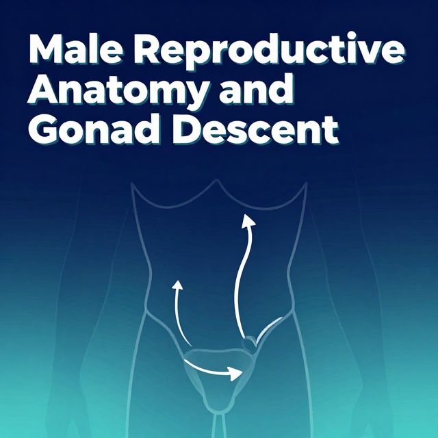
3.2. Testicular Descent
This is a complex process involving several steps:
- Gubernaculum Shortening: The gubernaculum shortens, pulling the testis caudally.
- Processus Vaginalis Formation: A pouch forms in the body wall called the 📚 processus vaginalis. The testis migrates dorsal to this pouch.
- Inguinal Canal Formation: As the testis and processus vaginalis descend, they push through the abdominal wall, taking layers of musculature and fascia with them, thereby creating the 📚 inguinal canal.
3.3. Inguinal Canal Layers
The layers of the abdominal wall contribute to the coverings of the spermatic cord as it passes through the inguinal canal:
- Parietal peritoneum → Tunica vaginalis
- Transversalis fascia → Internal spermatic fascia
- Transversus abdominis muscle → No direct contribution (it goes underneath)
- Internal oblique muscle → Cremaster fascia and muscle
- External oblique muscle → External spermatic fascia
3.4. Contents of the Inguinal Canal
- Male:
- Ductus deferens
- Testicular artery
- Pampiniform venous plexus
- Ilioinguinal nerve
- Genital branch of the genitofemoral nerve
- Remnants of the processus vaginalis
- Autonomic nerves and lymphatics
- Female:
- Round ligament of the uterus
- Ilioinguinal nerve
- Genital branch of the genitofemoral nerve
- Remnants of the processus vaginalis
- Autonomic nerves and lymphatics
3.5. Defects in Testicular Descent
- 📚 Communicating Hydrocele: Occurs if the processus vaginalis fails to obliterate after testicular descent, allowing extra peritoneal fluid to collect within it.
- 📚 Congenital Inguinal Hernia: Intestinal loops protrude through a patent processus vaginalis, typically an indirect type of inguinal hernia.
- ⚠️ 📚 Cryptorchidism: An undescended testis where one or both testes remain in the inguinal canal (most common location).
- More common in premature males.
- Can spontaneously descend within the first six months; otherwise, surgical correction is needed.
- Associated with significant risks: infertility, testicular torsion, hernias, and an increased risk of germ-cell tumors.
3.6. Ovarian Descent
This process is more rudimentary compared to testicular descent.
- The gubernaculum attaches to the future uterus.
- Its cranial portion develops into the 📚 ovarian ligament.
- Its caudal portion forms the 📚 round ligament of the uterus, which extends into the labia majora.
- The cranial suspensory ligament persists as the 📚 suspensory ligament of the ovary.