Lower Limb Muscle Anatomy: Origin, Insertion, Innervation, and Action - kapak
Bilim#anatomy#lower limb#muscles#iliopsoas

Lower Limb Muscle Anatomy: Origin, Insertion, Innervation, and Action

Explore the detailed anatomy of the lower limb muscles, including the iliopsoas, gluteal, medial thigh, anterior thigh, and posterior thigh muscle groups, covering their origins, insertions, innervations, and specific actions.

rana_lJanuary 19, 2026 ~26 dk toplam
01

Sesli Özet

10 dakika

Konuyu otobüste, koşarken, yolda dinleyerek öğren.

Sesli Özet

Lower Limb Muscle Anatomy: Origin, Insertion, Innervation, and Action

0:009:56
02

Flash Kartlar

25 kart

Karta tıklayarak çevir. ← → ile gez, ⎵ ile çevir.

1 / 25
Tüm kartları metin olarak gör
  1. 1. What two main muscles comprise the iliopsoas group, and what is their primary collective action?

    The iliopsoas group is primarily composed of the Psoas major and the Iliacus muscles. Their primary collective action is hip flexion, making them crucial for movements like lifting the leg or bending at the waist. They are considered key players in hip movement.

  2. 2. Describe the origin, insertion, and innervation of the Psoas major muscle.

    The Psoas major originates from the T12 to L4 vertebrae and associated intervertebral disks, with deeper fibers from the L1 to L5 costal processes. It inserts onto the lesser trochanter of the femur. This muscle is innervated by the lumbar plexus, specifically spinal nerves L1, L2, and L3.

  3. 3. Explain the actions of the Psoas major muscle, differentiating between when the femur is fixed and when it is free.

    When the femur is free, the Psoas major primarily performs hip flexion and external rotation. However, when the femur is fixed, its actions change. Unilateral contraction causes lateral flexion of the trunk, while bilateral contraction helps raise the trunk from a supine position, such as during a sit-up.

  4. 4. What are the origin, insertion, and innervation of the Iliacus muscle, and what are its primary actions?

    The Iliacus muscle originates from the iliac fossa. It then inserts onto the lesser trochanter of the femur, sharing an insertion point with the Psoas major. This muscle is innervated by the femoral nerve, specifically spinal nerves L2 and L3. Its primary actions are hip flexion and external rotation.

  5. 5. Identify the origin, insertion, and innervation of the Gluteus maximus muscle.

    The Gluteus maximus originates from the dorsal surface of the sacrum, the posterior part of the ilium, the thoracolumbar fascia, and the sacrotuberous ligament. Its upper fibers insert into the iliotibial tract, while its lower fibers insert into the gluteal tuberosity of the femur. It is innervated by the inferior gluteal nerve, from L5 to S2.

  6. 6. Describe the primary actions of the Gluteus maximus, including the specific contributions of its upper and lower fibers.

    The entire Gluteus maximus muscle is a powerful extensor and external rotator of the hip. Additionally, its upper fibers contribute to hip abduction, helping to move the leg away from the midline. Conversely, its lower fibers assist in hip adduction, drawing the leg towards the midline.

  7. 7. What are the origin, insertion, and innervation of the Gluteus medius muscle?

    The Gluteus medius originates from the gluteal surface of the ilium, located below the iliac crest. It then inserts onto the lateral surface of the greater trochanter of the femur. This muscle is innervated by the superior gluteal nerve, which arises from spinal nerves L4 to S1.

  8. 8. Explain the main actions of the Gluteus medius and how its anterior and posterior parts contribute differently.

    The Gluteus medius primarily abducts the hip and is crucial for stabilizing the pelvis in the coronal plane, preventing it from dropping on the unsupported side during walking. Its anterior part also aids in hip flexion and internal rotation, while the posterior part assists in hip extension and external rotation.

  9. 9. Where does the Gluteus minimus originate and insert, and what are its primary actions and innervation?

    The Gluteus minimus originates from the ilium, situated beneath the gluteus medius. It inserts onto the anterolateral surface of the greater trochanter of the femur. This muscle is innervated by the superior gluteal nerve, and its primary actions include hip abduction, flexion, and internal rotation.

  10. 10. What is the origin, insertion, and innervation of the Tensor fasciae latae, and what are its main actions?

    The Tensor fasciae latae originates from the anterior superior iliac spine. It inserts into the iliotibial tract, a thick band of fascia running down the lateral thigh. This muscle is innervated by the superior gluteal nerve. Its main actions include tensing the fascia lata and contributing to hip abduction, flexion, and internal rotation.

  11. 11. Name the deep muscles primarily responsible for external rotation of the hip joint, and state their general innervation.

    The deep muscles primarily responsible for external rotation of the hip joint include the Obturator internus, the Gemelli (superior and inferior), and the Quadratus femoris. These muscles generally originate from the pelvic surface of the sacrum or around the ischium and insert near the greater trochanter. They are all innervated by the sacral plexus, specifically L5 and S1.

  12. 12. What is the general functional classification of the medial thigh muscles?

    The medial thigh muscles are functionally classified as the adductor group of the hip. Their primary role is to adduct the hip, meaning they draw the thigh towards the midline of the body. They also contribute to other movements like flexion, extension, and rotation, and play a role in pelvic stability.

  13. 13. Describe the origin, insertion, innervation, and actions of the Pectineus muscle.

    The Pectineus muscle originates from the pecten pubis. It inserts onto the pectineal line and the proximal linea aspera of the femur. This muscle is uniquely innervated by both the femoral and obturator nerves (L2 and L3). Its actions include hip adduction, external rotation, slight flexion, and stabilizing the pelvis.

  14. 14. What are the origin, insertion, innervation, and main actions of the Adductor longus?

    The Adductor longus originates from the superior pubic ramus. It inserts onto the medial lip of the linea aspera in the middle third of the femur. This muscle is innervated by the obturator nerve, from L2 to L4. Its main actions are hip adduction and flexion, and it also contributes to stabilizing the pelvis.

  15. 15. Identify the origin, insertion, innervation, and primary actions of the Adductor brevis.

    The Adductor brevis originates from the inferior pubic ramus. It inserts onto the medial lip of the linea aspera. This muscle is innervated by the obturator nerve, specifically L2 and L3. Its primary actions are hip adduction and flexion, similar to the Adductor longus but often lying deeper.

  16. 16. What are the origin, insertion, innervation, and actions of the Gracilis muscle, including its knee actions?

    The Gracilis muscle originates from the inferior pubic ramus, below the pubic symphysis. It inserts into the medial border of the tibial tuberosity, forming part of the pes anserinus. Innervated by the obturator nerve (L2 and L3), it adducts the hip and also flexes and internally rotates the knee.

  17. 17. Describe the origin, insertion, innervation, and actions of the Obturator externus.

    The Obturator externus originates from the outer surface of the obturator membrane. It inserts into the trochanteric fossa of the femur. This muscle is innervated by the obturator nerve, specifically L3 and L4. Its actions include hip adduction and external rotation, and it also helps stabilize the pelvis.

  18. 18. Explain the origin, insertion, innervation, and actions of the Adductor magnus, noting the differences between its deep and superficial parts.

    The Adductor magnus is a large muscle originating from the inferior pubic ramus, ischial ramus, and ischial tuberosity. Its deep part inserts onto the medial lip of the linea aspera and is innervated by the obturator nerve (L2-L4). Its superficial part inserts onto the adductor tubercle of the femur and is innervated by the tibial nerve (L4). This powerful muscle performs hip adduction, extension, and slight flexion, with its tendinous insertion contributing to internal rotation and pelvic stabilization.

  19. 19. What are the origin, insertion, innervation, and actions of the Sartorius muscle, including both hip and knee movements?

    The Sartorius muscle originates from the anterior superior iliac spine. It inserts medially to the tibial tuberosity, as part of the pes anserinus. Innervated by the femoral nerve (L2 and L3), it flexes, abducts, and externally rotates the hip, while also flexing and internally rotating the knee. It's often called the "tailor's muscle" due to its actions.

  20. 20. Name the four muscles that comprise the Quadriceps femoris group and state their common insertion and primary collective action.

    The Quadriceps femoris group comprises the Rectus femoris, Vastus medialis, Vastus lateralis, and Vastus intermedius. All four muscles insert via the patellar ligament onto the tibial tuberosity. Their primary collective action is to extend the knee, making them essential for standing, walking, and running.

  21. 21. Which muscle of the Quadriceps femoris group is unique in its origin and additional action at the hip joint?

    The Rectus femoris is unique among the quadriceps muscles. It originates from the anterior inferior iliac spine and the acetabular roof of the hip joint. Unlike the other vasti muscles, which only cross the knee, the Rectus femoris also crosses the hip joint, allowing it to flex the hip in addition to extending the knee.

  22. 22. What are the general origins of the Vastus medialis, Vastus lateralis, and Vastus intermedius, and what is their primary action?

    The Vastus medialis, Vastus lateralis, and Vastus intermedius originate from various parts of the femoral shaft, including the linea aspera and intertrochanteric line. These three muscles form the bulk of the anterior thigh. Their primary action is knee extension, contributing significantly to the power of the quadriceps group.

  23. 23. Describe the Articularis genus muscle, including its origin, insertion, and specific function.

    The Articularis genus consists of distal fibers of the Vastus intermedius. It originates from the anterior side of the femoral shaft. It inserts into the suprapatellar recess of the knee joint capsule. Its specific function is to retract the suprapatellar bursa during knee extension, preventing it from becoming entrapped between the femur and the patella.

  24. 24. What is the common functional classification of the posterior thigh muscles, and what are their primary actions?

    The posterior thigh muscles are commonly known as the hamstrings. This group primarily functions to flex the knee and extend the hip. They are crucial for activities like walking, running, and jumping, and also play a significant role in stabilizing the pelvis.

  25. 25. Describe the origins, insertion, and innervation of the two heads of the Biceps femoris muscle.

    The Biceps femoris has two heads: the long head originates from the ischial tuberosity and sacrotuberous ligament, while the short head originates from the lateral lip of the linea aspera. Both heads converge to insert onto the head of the fibula. The long head is innervated by the tibial nerve, and the short head by the common fibular nerve, both from L5 to S2.

03

Bilgini Test Et

15 soru

Çoktan seçmeli sorularla öğrendiklerini ölç. Cevap + açıklama.

Soru 1 / 15Skor: 0

Which of the following muscles is composed of two main parts, the Psoas major and the Iliacus, and is a key player in hip flexion?

04

Detaylı Özet

6 dk okuma

Tüm konuyu derinlemesine, başlık başlık.

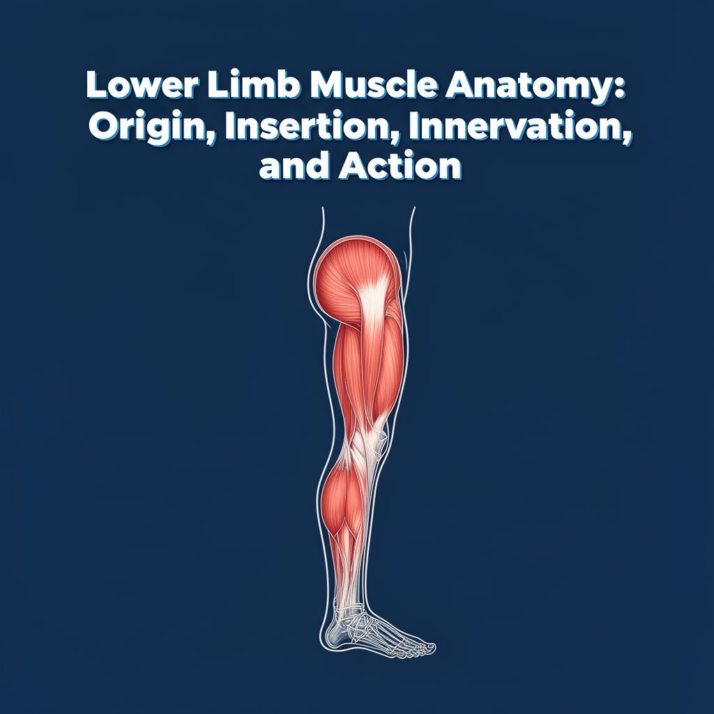
📚 Lower Limb Muscles: Anatomy and Function

This study material provides a comprehensive overview of the muscles of the lower limb, detailing their origins, insertions, innervations, and primary actions. It has been compiled and organized from a lecture audio transcript and supplementary copy-pasted text, including anatomical tables and diagrams.


🦵 Introduction to Lower Limb Muscles

The muscles of the lower limb are crucial for human movement, locomotion, stability, and maintaining posture. Understanding their individual roles and how they work together is fundamental to comprehending biomechanics and functional anatomy. This guide categorizes these muscles into key groups based on their anatomical location and primary functions.


1️⃣ Iliopsoas Group: Primary Hip Flexors

The iliopsoas group is a powerful hip flexor, essential for movements like walking and running.

📚 Key Muscles:

  • Psoas Major
    • Origin:
      • Superficial: T12-L4 vertebrae and associated intervertebral disks (lateral surfaces).
      • Deep: L1-L5 vertebrae (costal processes).
    • Insertion: Lesser trochanter of the femur.
    • Innervation: Lumbar plexus (L1, L2, L3).
    • Actions:
      • Hip Joint: Flexion and external rotation.
      • Lumbar Spine (with fixed femur): Unilateral contraction flexes the trunk laterally; bilateral contraction raises the trunk from a supine position.
  • Iliacus
    • Origin: Iliac fossa.
    • Insertion: Lesser trochanter of the femur.
    • Innervation: Femoral nerve (L2-L3).
    • Actions: Hip flexion and external rotation.

💡 Note on Psoas Minor: Present in approximately 50% of the population, it lies superficial to the Psoas major. However, it is not considered a lower limb muscle as its actions are confined to the abdomen.


2️⃣ Gluteal Muscles: Hip Movement and Stability

The gluteal muscles are vital for hip movement, stability, and maintaining posture, forming the bulk of the buttock region.

📚 Key Muscles:

  • Gluteus Maximus
    • Origin: Dorsal surface of the sacrum (lateral part), ilium (gluteal surface, posterior part), thoracolumbar fascia, sacrotuberous ligament.
    • Insertion: Upper fibers into the iliotibial tract; lower fibers into the gluteal tuberosity of the femur.
    • Innervation: Inferior gluteal nerve (L5-S2).
    • Actions:
      • Entire Muscle: Extends and externally rotates the hip.
      • Upper Fibers: Abduction.
      • Lower Fibers: Adduction.
  • Gluteus Medius
    • Origin: Ilium (gluteal surface below the iliac crest, between anterior and posterior gluteal lines).
    • Insertion: Lateral surface of the greater trochanter of the femur.
    • Innervation: Superior gluteal nerve (L4-S1).
    • Actions:
      • Entire Muscle: Abducts the hip, stabilizes the pelvis in the coronal plane.
      • Anterior Part: Flexion and internal rotation.
      • Posterior Part: Extension and external rotation.
  • Gluteus Minimus
    • Origin: Ilium (gluteal surface below the origin of gluteus medius).
    • Insertion: Anterolateral surface of the greater trochanter of the femur.
    • Innervation: Superior gluteal nerve (L4-S1).
    • Actions: Hip abduction, flexion, and internal rotation.
  • Tensor Fasciae Latae
    • Origin: Anterior superior iliac spine.
    • Insertion: Iliotibial tract.
    • Innervation: Superior gluteal nerve (L4-S1).
    • Actions: Tenses the fascia lata; hip abduction, flexion, and internal rotation.
  • Piriformis
    • Origin: Pelvic surface of the sacrum.
    • Insertion: Apex of the greater trochanter of the femur.
    • Innervation: Sacral plexus (S1, S2).
    • Actions: External rotation, abduction, and extension of the hip joint; stabilizes the hip joint.
  • Deep Gluteal Muscles (Obturator Internus, Gemelli, Quadratus Femoris)
    • These muscles primarily contribute to external rotation of the hip joint.
    • Obturator Internus: Originates from the inner surface of the obturator membrane.
    • Gemelli (Superior & Inferior): Originate from the ischial spine and ischial tuberosity, respectively.
    • Quadratus Femoris: Originates from the lateral border of the ischial tuberosity.
    • Insertion: All insert around the greater trochanter or intertrochanteric crest.
    • Innervation: Sacral plexus (L5, S1).

3️⃣ Medial Thigh Muscles: The Adductor Group

Functionally, these muscles are considered the adductors of the hip, responsible for bringing the thigh towards the midline. They are divided into superficial and deep layers.

📚 Superficial Layer:

  • Pectineus
    • Origin: Pecten pubis.
    • Insertion: Pectineal line and proximal linea aspera of the femur.
    • Innervation: Femoral nerve, obturator nerve (L2, L3).
    • Actions: Hip adduction, external rotation, and slight flexion; stabilizes the pelvis.
  • Adductor Longus
    • Origin: Superior pubic ramus.
    • Insertion: Linea aspera (medial lip in the middle third of the femur).
    • Innervation: Obturator nerve (L2-L4).
    • Actions: Hip adduction and flexion (up to 70 degrees); extension (past 80 degrees of flexion); stabilizes the pelvis.
  • Adductor Brevis
    • Origin: Inferior pubic ramus.
    • Insertion: Linea aspera.
    • Innervation: Obturator nerve (L2, L3).
    • Actions: Hip adduction and flexion.
  • Gracilis
    • Origin: Inferior pubic ramus below the pubic symphysis.
    • Insertion: Medial border of the tibial tuberosity (part of the pes anserinus).
    • Innervation: Obturator nerve (L2, L3).
    • Actions: Hip adduction; knee flexion and internal rotation.

📚 Deep Layer:

  • Obturator Externus
    • Origin: Outer surface of the obturator membrane and its bony boundaries.
    • Insertion: Trochanteric fossa of the femur.
    • Innervation: Obturator nerve (L3, L4).
    • Actions: Hip adduction and external rotation; stabilizes the pelvis.
  • Adductor Magnus
    • Origin: Inferior pubic ramus, ischial ramus, and ischial tuberosity.
    • Insertion:
      • Deep part ("fleshy insertion"): Medial lip of the linea aspera.
      • Superficial part ("tendinous insertion"): Adductor tubercle of the femur.
    • Innervation:
      • Deep part: Obturator nerve (L2-L4).
      • Superficial part: Tibial nerve (L4).
    • Actions: Hip adduction, extension, and slight flexion (tendinous insertion also active in internal rotation); stabilizes the pelvis.

4️⃣ Anterior Thigh Muscles: Knee Extensors

These muscles are primarily responsible for extending the knee joint.

📚 Key Muscles:

  • Sartorius
    • Origin: Anterior superior iliac spine.
    • Insertion: Medial to the tibial tuberosity (part of the pes anserinus).
    • Innervation: Femoral nerve (L2, L3).
    • Actions: Hip flexion, abduction, and external rotation; knee flexion and internal rotation.
  • Quadriceps Femoris Group
    • This group consists of four muscles that collectively extend the knee. All four muscles insert via the patellar ligament onto the tibial tuberosity.
    • Innervation (all): Femoral nerve (L2-L4).
    • Rectus Femoris
      • Origin: Anterior inferior iliac spine and acetabular roof of the hip joint.
      • Actions: Hip flexion and knee extension (unique among quadriceps for hip action).
    • Vastus Medialis
      • Origin: Linea aspera (medial lip), intertrochanteric line (distal part).
      • Actions: Knee extension.
    • Vastus Lateralis
      • Origin: Linea aspera (lateral lip), greater trochanter (lateral surface).
      • Actions: Knee extension.
    • Vastus Intermedius
      • Origin: Femoral shaft (anterior side).
      • Actions: Knee extension.
    • Articularis Genus: Distal fibers of the vastus intermedius.
      • Origin: Anterior side of femoral shaft at the level of the suprapatellar recess.
      • Insertion: Suprapatellar recess of knee joint capsule.
      • Actions: Retracts the suprapatellar bursa to prevent entrapment during knee extension.

5️⃣ Posterior Thigh Muscles: Knee Flexors (Hamstrings)

Commonly known as the hamstrings, these muscles primarily flex the knee and extend the hip.

📚 Key Muscles:

  • Biceps Femoris
    • Insertion (both heads): Head of fibula.
    • Long Head
      • Origin: Ischial tuberosity, sacrotuberous ligament (common head with semitendinosus).
      • Innervation: Tibial nerve (L5-S2).
      • Actions: Hip extension, stabilizes the pelvis in the sagittal plane; knee flexion and external rotation.
    • Short Head
      • Origin: Lateral lip of the linea aspera in the middle third of the femur.
      • Innervation: Common fibular nerve (L5-S2).
      • Actions: Knee flexion and external rotation.
  • Semimembranosus
    • Origin: Ischial tuberosity.
    • Insertion: Medial tibial condyle, oblique popliteal ligament, popliteus fascia.
    • Innervation: Tibial nerve (L5-S2).
    • Actions: Hip extension, stabilizes the pelvis in the sagittal plane; knee flexion and internal rotation.
  • Semitendinosus
    • Origin: Ischial tuberosity and sacrotuberous ligament (common head with long head of biceps femoris).
    • Insertion: Medial to the tibial tuberosity (part of the pes anserinus).
    • Innervation: Tibial nerve (L5-S2).
    • Actions: Hip extension, stabilizes the pelvis in the sagittal plane; knee flexion and internal rotation.

Kendi çalışma materyalini oluştur

PDF, YouTube videosu veya herhangi bir konuyu dakikalar içinde podcast, özet, flash kart ve quiz'e dönüştür. 1.000.000+ kullanıcı tercih ediyor.

Sıradaki Konular

Tümünü keşfet
Muscles of the Lower Limb: Leg Compartments

Muscles of the Lower Limb: Leg Compartments

Explore the anatomy of the leg muscles, categorized by their compartments: lateral, anterior, and posterior. Learn about the origin, insertion, innervation, and actions of key muscles.

Özet 25 15
Anatomy of Shoulder and Arm Muscles: A Detailed Guide

Anatomy of Shoulder and Arm Muscles: A Detailed Guide

Explore the detailed anatomy, origins, insertions, innervations, and actions of key muscles in the shoulder and arm regions, including anterior, posterior, and arm flexor/extensor groups.

Özet 24 15
The Musculoskeletal System: Structure, Function, and Locomotion

The Musculoskeletal System: Structure, Function, and Locomotion

Explore the intricate musculoskeletal system, its components, functions, and the mechanisms of locomotion in various organisms, with a detailed focus on the human body.

Özet 25 15 Görsel
Skeletal Muscle Structure and Neuromuscular Junction

Skeletal Muscle Structure and Neuromuscular Junction

This audio summary provides an academic overview of skeletal muscle terminology, morphology, filament and sarcomere structure, innervation, and the intricate process of excitation-contraction coupling.

7 dk Özet 18 12
Neuron Types and Nervous System Structures

Neuron Types and Nervous System Structures

This summary explores the diverse morphology and classification of neurons, their fundamental components, synaptic connections, and the crucial role of glial cells and peripheral nervous system structures.

6 dk Özet 25 15
Male Reproductive Anatomy and Gonad Descent

Male Reproductive Anatomy and Gonad Descent

Explore the gross anatomy of the male reproductive system, accessory glands, and the embryological development and descent of gonads, including clinical correlations.

7 dk Özet 15
The Visual Pathway, Reflexes, and Eye Movements

The Visual Pathway, Reflexes, and Eye Movements

An academic overview of the visual pathway from retina to cortex, including visual field processing, magnocellular and parvocellular streams, light and accommodation reflexes, and conjugate eye movements.

8 dk Özet 25 15
The Anatomy and Physiology of the Human Heart

The Anatomy and Physiology of the Human Heart

Explore the intricate structure and function of the human heart, covering its location, chambers, valves, and the electrical conduction system that drives its rhythmic beat.

Özet 25 15