The Musculoskeletal System: Structure, Function, and Locomotion - kapak
Bilim#musculoskeletal system#skeletal system#muscular system#locomotion

The Musculoskeletal System: Structure, Function, and Locomotion

Explore the intricate musculoskeletal system, its components, functions, and the mechanisms of locomotion in various organisms, with a detailed focus on the human body.

izemApril 26, 2026 ~17 dk toplam
01

Görsel Özet

İnfografik

Konunun tüm parçalarını tek bakışta gör.

The Musculoskeletal System: Structure, Function, and Locomotion - görsel özet infografik
Tam boyutta görüntüle →
02

Flash Kartlar

25 kart

Karta tıklayarak çevir. ← → ile gez, ⎵ ile çevir.

1 / 25
Tüm kartları metin olarak gör
  1. 1. What is the primary function of the musculoskeletal system?

    The primary function of the musculoskeletal system is locomotion, which is the capacity to move independently from one place to another. It also provides support, protection for organs, and enables various body movements through the coordinated action of bones, joints, and muscles.

  2. 2. Why is locomotion vital for the survival of organisms?

    Locomotion is vital for survival because it allows organisms to perform essential activities such as finding food, locating suitable habitats, escaping from danger, and successfully reproducing. Without the ability to move, many organisms would struggle to meet their basic needs and perpetuate their species.

  3. 3. Describe how single-celled organisms achieve locomotion.

    Single-celled organisms achieve locomotion using specialized structures. For example, some use pseudopods, which are temporary projections of the cell membrane that allow them to 'crawl.' Others use cilia, which are short, hair-like structures that beat rhythmically to propel the organism through its environment.

  4. 4. What is the fundamental principle underlying all forms of locomotion?

    The fundamental principle underlying all forms of locomotion involves contractile proteins. These proteins have the unique ability to change length, causing movement. This mechanism is observed across various organisms, from the simplest single-celled entities to complex multicellular animals, enabling diverse forms of movement.

  5. 5. How does an earthworm achieve movement?

    An earthworm achieves movement by utilizing a combination of circular and longitudinal muscles working against a fluid skeleton. The contraction of circular muscles elongates the body, while longitudinal muscles shorten it. This coordinated action, coupled with anchoring bristles, allows it to extend and contract, resulting in a crawling motion.

  6. 6. What percentage of the human body weight does the musculoskeletal system comprise?

    The human musculoskeletal system comprises approximately 20% of the total body weight. This significant portion highlights its crucial role in supporting the body, enabling movement, and protecting vital organs, making it one of the largest and most essential systems in the body.

  7. 7. How many bones are typically found in the adult human body?

    The adult human body typically contains 206 bones. These bones form the skeletal framework, providing structure, support, and protection for internal organs, and serving as attachment points for muscles, which are essential for movement.

  8. 8. What is the primary function of ligaments in the human body?

    The primary function of ligaments is to connect bones to other bones. They are strong, fibrous connective tissues that provide stability to joints, preventing excessive or unwanted movements. Ligaments are crucial for maintaining the structural integrity and proper alignment of the skeletal system.

  9. 9. What is the primary function of tendons in the human body?

    The primary function of tendons is to attach muscles to bones. They are tough, flexible bands of fibrous connective tissue that transmit the force generated by muscle contraction to the bones. This transmission of force is what enables movement of the skeletal system.

  10. 10. What are the main functions of bones in the human skeletal system?

    Bones in the human skeletal system serve several vital functions. They provide attachment sites and levers for muscles, enabling movement, offer body shape and support, protect delicate organs like the brain and heart, and store essential minerals such as calcium and phosphorus.

  11. 11. Describe the nature of bone tissue.

    Bone tissue is a highly active, living tissue that is constantly remodeling. It undergoes continuous processes of formation by osteoblasts and reabsorption by osteoclasts, allowing it to adapt to stress, repair damage, and maintain mineral homeostasis. This dynamic nature makes bone a responsive and resilient tissue.

  12. 12. What is an osteon and what does it contain?

    An osteon is the basic microscopic unit of compact bone, appearing as a cylindrical structure. It surrounds a central Haversian canal, which contains blood vessels and nerves that supply the bone tissue. Osteons are arranged in parallel, providing strength and efficient nutrient delivery to the dense bone matrix.

  13. 13. What is the role of the periosteum?

    The periosteum is a tough outer membrane that covers the surface of bones, except at articular surfaces. It is crucial for new bone production, especially during growth and repair after injury. Additionally, it serves as an important attachment site for muscles and tendons, facilitating their connection to the bone.

  14. 14. Name the three main types of bone cells and their respective functions.

    The three main types of bone cells are osteoblasts, which are responsible for bone formation by synthesizing new bone matrix; osteocytes, which are mature bone cells that maintain the bone matrix; and osteoclasts, which are large cells involved in bone reabsorption and remodeling by breaking down bone tissue.

  15. 15. Differentiate between compact bone and spongy bone.

    Compact bone is dense and solid, forming the outer layer of most bones, providing strength and protection. Spongy bone, also known as cancellous bone, is porous with a network of bony plates called trabeculae, and its spaces are filled with bone marrow. Spongy bone is found at the ends of long bones and in the interior of other bones.

  16. 16. What is the difference between red marrow and yellow marrow?

    Red marrow is found in the spaces of spongy bone and is responsible for producing blood cells (hematopoiesis), including red blood cells, white blood cells, and platelets. Yellow marrow, primarily composed of fat cells, is found in the medullary cavity of long bones and can convert to red marrow if the body requires increased blood cell production.

  17. 17. List the five classifications of bones based on their shape.

    Bones are classified into five types based on their shape: long bones (e.g., femur), short bones (e.g., carpals), flat bones (e.g., skull bones), irregular bones (e.g., vertebrae), and sesamoid bones (e.g., patella). Each shape contributes to specific functions within the skeletal system.

  18. 18. What is the role of cartilage in the skeletal system, both during development and in adult joints?

    During development, cartilage gradually ossifies into bone, forming much of the skeleton. In adult joints, cartilage provides support, reduces friction between bones, and acts as a shock absorber, cushioning the ends of bones and facilitating smooth, pain-free movement. It is a flexible connective tissue.

  19. 19. What are the two main divisions of the human skeleton? Provide examples for each.

    The two main divisions of the human skeleton are the axial skeleton, which includes the skull, vertebrae, ribs, and sternum, forming the central axis of the body. The appendicular skeleton comprises the arms, legs, and the pectoral and pelvic girdles, which attach the limbs to the axial skeleton.

  20. 20. Describe the characteristics of immovable fibrous joints and give an example.

    Immovable fibrous joints, also known as synarthroses, are characterized by bones held together by dense fibrous connective tissue, allowing little to no movement. These joints provide strong connections and protection. A prime example of such a joint is the sutures found between the bones of the cranium.

  21. 21. Name and describe two types of highly mobile synovial joints.

    Two types of highly mobile synovial joints are ball-and-socket joints, like the shoulder, which allow movement in multiple planes (flexion, extension, abduction, adduction, rotation). Hinge joints, like the elbow, primarily allow movement in one plane (flexion and extension), similar to a door hinge.

  22. 22. What is the function of synovial fluid in joints?

    Synovial fluid is a viscous, egg-white-like fluid found in the cavities of synovial joints. Its primary function is to lubricate the joint, reducing friction between the articular cartilages during movement. It also provides nutrients to the avascular cartilage and removes waste products, contributing to joint health.

  23. 23. What gives muscle fibers their striated appearance?

    Muscle fibers appear striated due to the organized arrangement of myofilaments within their myofibrils. Specifically, the alternating pattern of thick myosin and thin actin filaments, which are organized into repeating functional subunits called sarcomeres, creates this characteristic striped or banded look under a microscope.

  24. 24. Explain the basic principle of muscle contraction according to the sliding filament theory.

    According to the sliding filament theory, muscle contraction occurs when actin and myosin filaments slide past each other. Myosin heads bind to actin, form cross-bridges, and then pivot, pulling the actin filaments towards the center of the sarcomere. This shortens the sarcomere, and consequently the entire muscle fiber, without the filaments themselves changing length.

  25. 25. What is the primary energy source for muscle contraction?

    The primary energy source for muscle contraction is Adenosine Triphosphate (ATP). Mitochondria within muscle cells produce ATP through cellular respiration. This ATP powers the binding and unbinding of myosin heads to actin, driving the sliding filament mechanism and enabling the muscle to contract and relax.

03

Bilgini Test Et

15 soru

Çoktan seçmeli sorularla öğrendiklerini ölç. Cevap + açıklama.

Soru 1 / 15Skor: 0

What is the primary function of the musculoskeletal system as described in the text?

04

Detaylı Özet

7 dk okuma

Tüm konuyu derinlemesine, başlık başlık.

This study material has been compiled from various sources, including lecture audio transcripts and copy-pasted text provided by the user.


🦴💪 The Musculoskeletal System: Structure, Function, and Locomotion

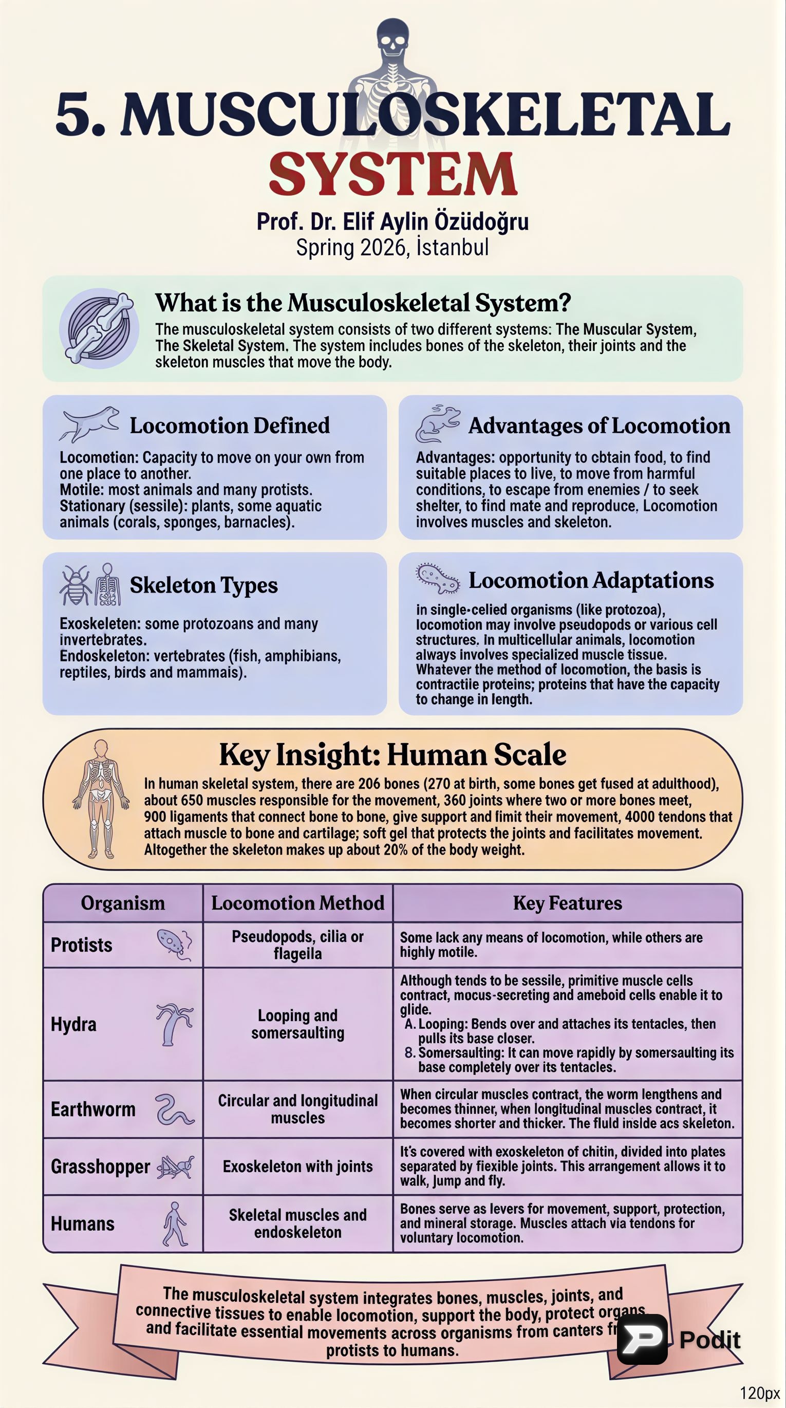
The musculoskeletal system is a complex and integrated biological system that provides form, support, stability, and movement to the body. It is fundamentally composed of two distinct but interconnected systems: the Skeletal System and the Muscular System. Together, these systems enable locomotion and perform numerous vital functions for survival.

🚶‍♀️ Locomotion: The Power of Movement

Locomotion is defined as the capacity of an organism to move independently from one place to another. It is a critical aspect of survival for most living beings.

✅ Importance of Locomotion

  • Obtaining Food: Allows organisms to hunt or forage.
  • Finding Suitable Habitats: Enables migration to favorable environments.
  • Escaping Harmful Conditions: Facilitates avoidance of danger.
  • Evading Predators/Seeking Shelter: Provides protection.
  • Finding Mates and Reproduction: Essential for species propagation.

🌍 Diversity of Locomotion in Organisms

While some organisms are stationary (sessile), such as plants and certain aquatic animals (e.g., corals, sponges, barnacles), most animals and many protists are motile.

  • Basic Principle: Whatever the method, locomotion is fundamentally based on contractile proteins that can change in length.
  • Single-celled Organisms (Protozoa): May use specialized cell structures like pseudopods, cilia, or flagella.
  • Multicellular Animals: Always involve specialized muscle tissue.
  • Exoskeleton vs. Endoskeleton:
    • 📚 Exoskeleton: A rigid external covering, found in some protozoans and many invertebrates (e.g., grasshoppers).
    • 📚 Endoskeleton: An internal support structure, characteristic of vertebrates (e.g., fish, amphibians, reptiles, birds, and mammals).

🔬 Examples of Locomotion Mechanisms

  • Hydra: Although generally sessile, hydras can move through:
    • Looping: Bending over to attach tentacles, then pulling the base closer.
    • Somersaulting: Rapid movement by flipping the base completely over the tentacles.
  • Earthworm: Utilizes a hydrostatic skeleton.
    • When circular muscles contract, the worm lengthens and thins.
    • When longitudinal muscles contract, it shortens and thickens.
    • The fluid inside acts as a skeleton, providing resistance for muscle action.
  • Grasshopper: Possesses a chitinous exoskeleton divided into plates with flexible joints, allowing it to walk, jump, and fly.

🧍 The Human Musculoskeletal System: An Overview

The human musculoskeletal system is a marvel of biological engineering, making up approximately 20% of body weight.

📊 Key Components and Numbers

  • Bones: 206 (adults; 270 at birth, some fuse later)
  • Muscles: ~650 responsible for movement
  • Joints: 360, where two or more bones meet
  • Ligaments: 900, tough elastic tissues connecting bone to bone, providing support and limiting movement.
  • Tendons: 4000, fibrous connective tissues attaching muscle to bone.
  • Cartilage: Soft gel-like tissue protecting joints and facilitating movement.

🦴 The Skeletal System: Support and Protection

The skeletal system is composed of bones, cartilage, ligaments, and other connective tissues.

✅ Functions of Bones

  1. Muscle Attachment & Levers: Serve as sites for skeletal muscle attachment and act as levers to produce movement when muscles contract.
  2. Shape & Support: Give the body its characteristic shape and support body structures.
  3. Protection: Safeguard delicate internal organs (e.g., brain, spinal cord, heart, lungs).
  4. Mineral Storage: Act as a reservoir for essential minerals like calcium and phosphorus.

🔬 Bone Structure and Composition

Bone is a highly active, living tissue, constantly undergoing remodeling through absorption of old tissue and formation of new tissue.

  • Composition: Made of living cells, connective tissue fibers, and inorganic compounds.
  • Microscopic Unit: The basic microscopic unit is the osteon (Haversian system).
    • Osteons are cylindrical structures (~0.2 mm) consisting of concentric lamellae of compact bone tissue.
    • They surround a central (Haversian) canal which contains blood vessels and nerves.
  • Periosteum: A tough membrane covering the outer surface of bones (except at joints).
    • Functions: Production of new bone for growth and repair, and serves as an attachment point for muscles.

🧬 Bone Cells

There are three primary types of bone cells:

  1. Osteoblasts: 📚 Bone-forming cells responsible for synthesizing and mineralizing bone matrix (osteoid).
  2. Osteocytes: 📚 Mature bone cells, derived from osteoblasts, that maintain the mineral concentration of the bone matrix. They are the longest-living bone cells (90-95% of bone cells).
  3. Osteoclasts: 📚 Specialized, multinucleated giant cells that reabsorb bone tissue, crucial for bone remodeling and development.

🔄 Bone Formation (Ossification)

Osteoblasts secrete collagen molecules and polysaccharides. Collagen fibers bind with polysaccharides to form a cement-like substance. Calcium and phosphate ions from body fluids then combine to form calcium phosphate, which precipitates within these fibers and cement, leading to bone hardening.

🦴 Types of Bony Tissue

Most bones contain both types:

  • Compact Bone: Dense and strong, forming the outer layer of bones.
  • Spongy Bone (Cancellous Bone): More porous, found inside bones.
    • Spaces within spongy bone are filled with marrow.
    • Red Marrow: Found in vertebrae, ribs, breastbone, cranium, and long bones; produces red blood cells, platelets, and some white blood cells.
    • Yellow Marrow: Primarily consists of fat cells, found in the long bones.

📏 Bone Shapes

Bones come in various sizes and shapes:

  • Long Bones (e.g., femur, humerus)
  • Short Bones (e.g., carpals, tarsals)
  • Flat Bones (e.g., skull, sternum)
  • Irregular Bones (e.g., vertebrae, facial bones)
  • Sesamoid Bones (e.g., patella)

🦵 Cartilage

Cartilage is a type of connective tissue that is more flexible than bone.

  • Development: In embryos, cartilage gradually ossifies (changes into bone) as minerals are deposited.
  • Adults: Found at the ends of ribs, in joints, the nose, and outer ear.
  • Functions: Provides support, flexibility, and cushions against impacts.

🧍 The Human Skeleton: Divisions

The human skeleton is divided into two main parts:

  1. Axial Skeleton: Forms the central axis of the body.
    • Includes the skull (cranium, facial, and jaw bones), vertebral column (33 vertebrae), ribs, and breastbone (sternum).
  2. Appendicular Skeleton: Comprises the limbs and the girdles that attach them to the axial skeleton.
    • Includes the arms, legs, pectoral girdle (shoulder blades and collar bones, connecting arms to the spine), and pelvic girdle (hip bones and pelvic bones, connecting legs to the spine).

🤝 Joints: Connections for Movement

Joints are points where two or more bones meet.

  • Immovable (Fibrous) Joints: Bones are tightly fitted together, allowing no movement (e.g., sutures of the cranium).
  • Movable Joints: Allow varying degrees of movement.
    • Hinge Joints: Allow back-and-forth motion (e.g., elbow, knee).
    • Ball-and-Socket Joints: Provide the widest range of movement (e.g., shoulder, hip).
    • Pivot Joints: Allow rotation (e.g., between the first two cervical vertebrae, allowing head rotation).
    • Gliding Joints: Allow limited flexibility and sliding movements (e.g., between vertebrae).

🔗 Ligaments and Tendons

  • Ligaments: Tough, fibrous bands of connective tissue that hold bones together at movable joints.
  • Synovial Fluid: Secreted into movable joints by surrounding membranes, acting as a lubricant to reduce friction.
  • Tendons: Strong fibers of connective tissue that attach muscles to bones.

💪 The Muscular System: The Engine of Movement

The muscular system is responsible for all body movements, both voluntary and involuntary.

🔬 Muscle Tissue Structure

Muscular tissue is composed of muscle fibers.

  • Each muscle fiber contains thousands of myofibrils.
  • Myofibrils are bundles of smaller protein filaments called myofilaments.
    • Thick filaments: Composed of the protein myosin.
    • Thin filaments: Composed of the protein actin.
  • These filaments are arranged in an overlapping pattern, giving muscle fibers a striped or striated appearance.
  • Myofibrils are divided into functional subunits called sarcomeres, which are the basic contractile units of muscle.

⚡ Muscle Contraction: The Sliding Filament Theory

According to the sliding filament theory, muscle fibers shorten when the actin and myosin filaments slide past one another.

  • This sliding action increases the overlap between the two types of filaments, thereby shortening the sarcomere and the entire muscle fiber.
  • The energy for this sliding process is supplied by ATP, which is produced by the mitochondria within muscle fibers.

🦵 Types of Muscle Tissue

There are three main types of muscle tissue, each with distinct characteristics and functions:
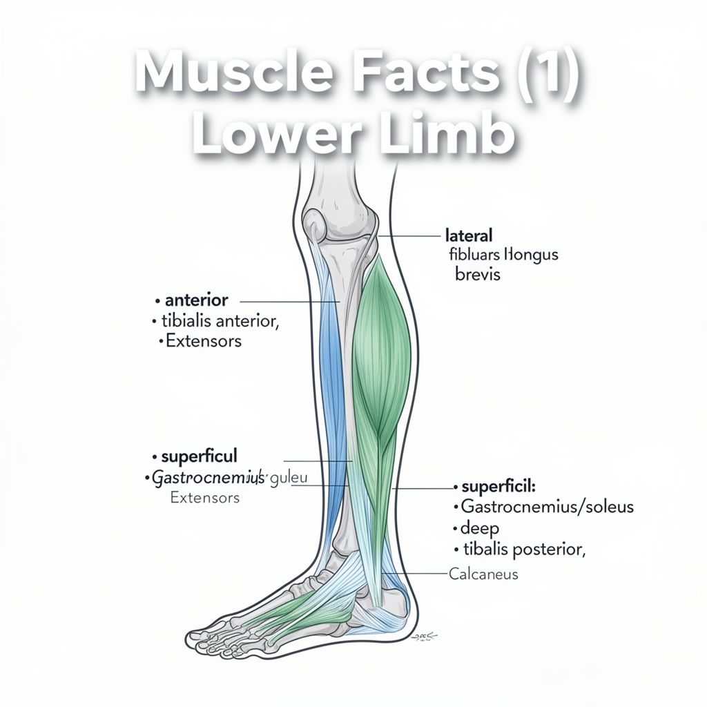
  1. Skeletal Muscle

    • Location: Attached to the bones of the skeleton by tendons.
    • Control: Responsible for all voluntary movements and locomotion.
    • Function: Works in antagonistic pairs (e.g., biceps and triceps) because muscles can only pull when they contract, not push.
    • Characteristics: Always maintains a slight state of contraction, known as muscle tone, even when relaxed.
    • Coordination: All voluntary movements are initiated and coordinated by impulses from the brain and spinal cord.
  2. Smooth Muscle

    • Location: Found in the walls of internal organs such as the digestive tract, blood vessels, and diaphragm.
    • Control: Operates without conscious control (involuntary).
    • Function: Responsible for processes like peristalsis (movement of food), blood pressure regulation, and other autonomic functions.
  3. Cardiac Muscle

    • Location: Found exclusively in the walls of the heart.
    • Control: Operates involuntarily.
    • Function: Responsible for pumping blood throughout the body.
    • Characteristics: Shares some structural features with skeletal muscle (striated) but functions like smooth muscle (involuntary).

This intricate system of bones, joints, and muscles works in harmony, enabling the vast array of movements essential for life and interaction with the environment.

Kendi çalışma materyalini oluştur

PDF, YouTube videosu veya herhangi bir konuyu dakikalar içinde podcast, özet, flash kart ve quiz'e dönüştür. 1.000.000+ kullanıcı tercih ediyor.

Sıradaki Konular

Tümünü keşfet
Bone Tissue and Joint Anatomy

Bone Tissue and Joint Anatomy

An academic summary detailing the composition, structure, cellular components, development, and pathologies of bone tissue, alongside an overview of joint types and synovial joint anatomy.

14 dk Özet 25 15
Muscles of the Lower Limb: Leg Compartments

Muscles of the Lower Limb: Leg Compartments

Explore the anatomy of the leg muscles, categorized by their compartments: lateral, anterior, and posterior. Learn about the origin, insertion, innervation, and actions of key muscles.

Özet 25 15
Bone Tissue and Joint Anatomy: A Comprehensive Overview

Bone Tissue and Joint Anatomy: A Comprehensive Overview

This summary provides an academic overview of bone tissue composition, types, cellular components, formation, degradation, associated diseases, and the anatomical classification and structure of joints.

12 dk Özet 25 15
Mastering Your Upper Limb Practical Exam

Mastering Your Upper Limb Practical Exam

Feeling nervous about your upper limb practical exam? This podcast breaks down the essential anatomy you need to know, from bones and muscles to nerves and vessels, to help you ace it!

Özet 23 10
Understanding Connective Tissues and Their Cells

Understanding Connective Tissues and Their Cells

Explore the fundamental types, structures, and cellular components of connective tissues, including their crucial roles in the body's support, protection, and immunity.

Özet 25 15
Nervous System: Structure, Function, and Signaling

Nervous System: Structure, Function, and Signaling

An academic overview of the nervous system's anatomical and functional divisions, cellular components, electrical signaling mechanisms, and inter-neuronal communication.

5 dk Özet 25 15
The Evolution of Science, Physiology, and Homeostasis

The Evolution of Science, Physiology, and Homeostasis

This summary explores the historical development of scientific thought, the emergence of physiology, and fundamental biological principles such as homeostasis, feedback mechanisms, and bioelectricity.

7 dk Özet 25 15
Fundamental Principles of Biological Organization and Nutrition

Fundamental Principles of Biological Organization and Nutrition

This summary explores the foundational concepts of biological organization, from cells to organ systems, and delves into essential nutritional components and their biochemical identification methods.

5 dk Özet 25 15