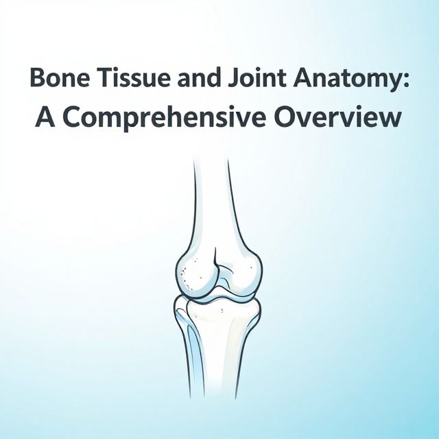
📚 Bone Tissue and Joints: A Comprehensive Study Guide
Source Information: This study material has been compiled and organized from a combination of copy-pasted text and a lecture audio transcript.
📝 Introduction to Bone Tissue and Joints
Bone tissue, a dynamic and essential component of the skeletal system, is characterized by its unique composition, structural organization, and specialized cellular elements. It exists in two primary forms: spongy and compact bone. The formation and remodeling of bone are complex processes, involving distinct cellular lineages and ossification mechanisms. Furthermore, the skeletal system incorporates various joint types, with synovial joints exhibiting intricate structures crucial for mobility and function. This guide will delineate these fundamental aspects, from the molecular constituents of bone to the macroscopic organization of joints and associated pathologies.
🦴 Bone Tissue Composition
Bone tissue is a composite material, consisting of both inorganic and organic components.
-
Inorganic Component (Approximately 65%)
- ✅ Primarily calcium phosphate in the form of pin-shaped hydroxyapatite crystals.
- ✅ Also includes water, bicarbonate, citrate, magnesium, sodium, and potassium.
- This component provides bone with its hardness and rigidity.
-
Organic Component (Approximately 35%)
- ✅ Composed of fibers and ground substance.
- ✅ Type I collagen constitutes 95% of the organic component, providing tensile strength and flexibility.
- ✅ Ground substance contains glycosaminoglycans (GAGs) and proteoglycans.
- ✅ Key glycoproteins include osteonectin, osteocalcin, osteopontin, and bone sialoprotein, which play roles in mineralization and cell adhesion.
🏗️ Bone Types Due to Structure (Secondary Bone Tissue)
Bone tissue is structurally classified into two main types:
-
Spongy Bone (Cancellous Bone)
- Found in the metaphysis and epiphysis of short and long bones, and in squamous bones.
- Characterized by a network of bone trabeculae (spiculae), which are not highly organized.
- Contains parallel lamellae.
- Bone marrow is located between the trabeculae.
- Bone cells acquire nutrients from vessels within the bone marrow via cytoplasmic extensions.
-
Compact Bone
-
Forms the external aspects of all bones, providing strength and protection.
-
Highly organized, containing canals, osteons, and other lamellar structures.
-
Canal Types:
- Haversian canals: Run parallel to the longitudinal axis of the bone.
- Volkmann canals: Connect Haversian canals to each other and to the periosteum and bone marrow.
- Vessels within these canals are surrounded by loose connective tissue containing nerve fibers.
-
Types of Lamellae:
- Special Lamellae (Osteons or Haversian System): Concentric rings of bone matrix around a central Haversian canal.
- Circumferential Lamellae:
- Inner Circumferential Lamellae: Line the inner surface of the compact bone.
- Outer Circumferential Lamellae: Located just beneath the periosteum.
- Interstitial Lamellae: Irregular fragments of old osteons found between intact osteons and circumferential lamellae.
-
🔬 Cellular Components and Bone Dynamics
Bone tissue is maintained by a dynamic interplay of specialized cells:
-
Osteoprogenitor Cells (Osteogenic Cells)
- 📚 Mesenchymal cells committed to becoming bone cells.
- Fusiform (spindle-shaped) like fibroblasts.
- Capable of mitosis and proliferation.
- Differentiate into osteoblasts under physiological conditions (e.g., osteon degeneration-regeneration) or pathological conditions (e.g., bone fracture).
- Can differentiate into chondrogenic cells at low oxygen levels.
- Contain few rough endoplasmic reticulum (GER), underdeveloped Golgi complex, and abundant free ribosomes.
- Localization: Inner aspect of the periosteum, connective tissue of Haversian and Volkmann canals, and endosteum.
-
Bone-Lining Cells
- An intermediate cell type between osteoprogenitor cells and osteoblasts.
- Similar properties to osteoprogenitor cells but cannot divide.
- Silent cells residing on the bone surface.
- Activate by appropriate signals to produce matrix.
- Possess gap junctions (nexuses) between them.
-
Osteoblasts
- Differentiate from osteoprogenitor cells, influenced by Bone Morphogenetic Protein (BMP) and Transforming Growth Factor-beta (TGF-β).
- Align in a single row, are polarized, and cuboidal or oval-shaped depending on activity.
- Possess gap junctions (nexuses) with each other and with osteocytes.
- Function: Produce the organic component of the bone matrix.
- Characterized by well-developed GER and Golgi complex, abundant secretory granules (e.g., alkaline phosphatase, pyrophosphatase), and a euchromatic nucleus.
- Products: Type I collagen, osteocalcin (mineralization), osteonectin (mineralization), osteopontin (forms sealing zone for osteoclasts), sialoprotein (binder to extracellular matrix), osteoprotegerin, M-CSF (macrophage-colony stimulating factor), RANKL (ligand for receptor activation of nuclear factor kappa B), receptor for parathyroid hormone, and osteoclast-stimulating factor.
- Differentiation into Osteocyte: When osteoblasts become entrapped within the uncalcified bone matrix (osteoid) they produced, and the tissue calcifies, their activity decreases, and they transform into osteocytes.
-
Osteocytes
- 📚 Mature bone cells residing within oval/squamous-shaped lacunae.
- Randomly oriented in primary bone, but oriented between neighboring lamellae in secondary bone.
- Canaliculi extend from lacunae, containing cytoplasmic extensions that connect osteocytes via gap junctions.
- Possess a heterochromatic nucleus and are poor in organelles compared to osteoblasts (decreased GER and Golgi).
- Nutrition is provided via cytoplasmic extensions.
- Cytoplasmic extensions shorten and connections are lost during aging.
- ⚠️ If osteocytes die, the surrounding matrix degenerates and is resorbed by osteoclasts.
-
Osteoclasts
- 📚 Large, multinuclear cells originating from the fusion of monocytes.
- Function: Degrade bone tissue (bone resorption).
- Reside at the surface of trabeculae or the inner aspect of compact bone, often in depressions called Howship lacunae.
- Control blood calcium levels.
- Acidophilic cytoplasm, cytoplasmic extensions at the bone tissue aspect.
- Rich in mitochondria and possess a well-developed Golgi.
- Contain lysosomal enzymes (e.g., collagenase, acid phosphatase).
- Howship Lacuna Structure:
- Ruffled border: Area where degradation takes place.
- Clear zone: Immediately neighbors the ruffled border, free of organelles but rich in actin.
- Vesicular zone: Contains endo- and exocytotic vesicles.
- Basal zone: Farthest from the Howship lacuna, containing organelles and nuclei.
- Sealing zone: Formed by the binding of osteopontin (from bone) and actin (from osteoclast) via integrins, creating a sealed compartment for degradation.
- Bone Degradation Mechanism:
- Osteoclasts possess carbonic anhydrase, which produces carbonic acid.
- Carbonic acid dissociates into hydrogen (H+) and bicarbonate (HCO3-) ions.
- A proton pump in the osteoclast membrane actively pumps H+ ions into the sealed compartment, decreasing the pH.
- The acidic environment degrades the inorganic component of the matrix.
- Lysosomal enzymes degrade the organic components.
- Tissue fragments are endocytosed, further degraded, and then expelled into the nearest capillary.
🛡️ Bone Sheaths
Bone is covered by two protective sheaths:
-
Periosteum
- Outer covering of bone.
- Outer fibrous layer: Composed of irregular dense connective tissue.
- Inner cellular layer: Contains osteoprogenitor cells.
- In mature bone, Sharpey fibers (collagen fibers) originate from the outer fibrous layer and extend into the outer circumferential and outer interstitial lamellae, anchoring the periosteum to the bone. These fibers are abundant at tendon-ligament attachments.
-
Endosteum
- Lines the inner aspect of compact bone, Haversian canals, and the surfaces of spongy bone trabeculae.
- Composed of reticular connective tissue.
- Contains osteoprogenitor cells.
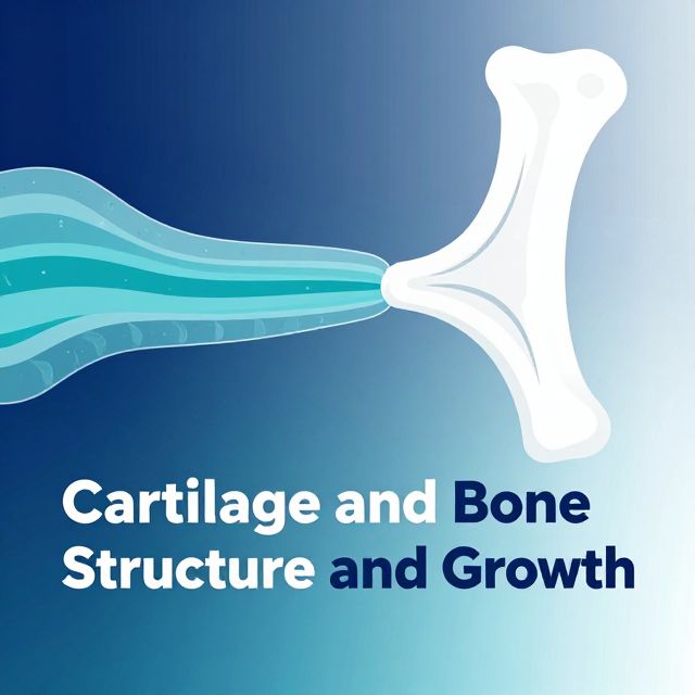
📈 Osteogenesis (Bone Development)
Bone development occurs through two primary mechanisms:
-
Intramembranous Ossification
- 📚 Direct differentiation of mesenchymal tissue into bone tissue.
- Forms the compact parts of short and long bones (e.g., flat bones of the skull).
- Process:
- Mesenchymal cells differentiate into osteoprogenitor cells, then into osteoblasts.
- Osteoblasts produce bone matrix (osteoid).
- Capillaries carry calcium and phosphorus into the osteoid, leading to calcification.
- Primary bone is formed, which is then organized into secondary bone tissue.
- Initially, a bony collar forms at the periphery of the diaphysis, inhibiting chondrocyte nourishment and causing degeneration.
- Osteoclasts pierce the bony collar to create nutritive pores.
- Periosteal vessels and osteoprogenitor cells migrate into the bone-forming area.
- Bone trabeculae with cores of calcified cartilage matrix are initially formed.
- Migrated mesenchymal and hematopoietic elements differentiate into bone marrow.
-
Endochondral Ossification
- 📚 Bone tissue replaces a pre-existing hyaline cartilage model.
- Forms the spongy parts of short and long bones.
- Process:
- A hyaline cartilage model forms at the interface of the epiphysis and metaphysis.
- Ions carried by vessels provide calcification of the matrix, forming the Primary Ossification Center at the diaphysis.
- As ossification proceeds, Secondary Ossification Centers appear at the epiphysis, with progenitor cells reaching this region.
- The cartilage model between the primary and secondary ossification centers is called the Epiphyseal Plate.
- Chondrocytes divide by mitosis, providing continuous lengthening of the cartilage model.
- The bony collar thickens and lengthens, progressively surrounding the cartilage, blocking nutrition, and promoting further bone formation.
- The epiphyseal plate is typically lost around age 20, marking the cessation of longitudinal growth.
- Bone Thickening (Appositional Growth):
- Occurs via appositional growth.
- Osteoprogenitor cells of the periosteum differentiate into osteoblasts.
- Newly formed bone tissue is added to the former one at the subperiosteal zone.
- The inner aspect of the bone tissue is eroded to enlarge the bone marrow cavity.
📊 Bone Regulation and Diseases
Bone development and remodeling are controlled by various factors and hormones.
-
Substances Decreasing Bone Degradation:
- Calcium, Vitamin D, Estrogen, Calcitonin.
- Biphosphonates: Decrease osteoclast number and activity.
-
Substances Increasing Bone Formation:
- Fluoride, Androgen, Growth Hormone.
- Statins: Stimulate differentiation into osteoblasts.
-
Bone Diseases:
- Osteoporosis: Loss of bone mass, increase in osteoclast number.
- Osteomalacia: Softening of bone due to defective mineralization of osteoid. Seen in Vitamin D insufficiency, renal-intestinal disease.
- Rickets: Mineralization defect at the epiphyseal plate (in children).
- Osteopetrosis: Dysfunction of osteoclasts, leading to abnormally dense but brittle bones.
- Osteosclerosis: Hyperactivity of osteoblasts, leading to increased bone density.
🤸 Joint Types
Joints are articulations between bones, classified by their structure and degree of movement.
-
Synarthrosis (Limited to No Movement)
- Fibrous: Bones joined by fibrous connective tissue.
- Examples: Tooth-alveolar (gomphosis), radioulnar (syndesmosis), tibiofibular (syndesmosis).
- Cartilaginous (Synchondrosis, Symphysis): Bones joined by cartilage.
- Examples: Vertebral (symphysis), sternocostal (synchondrosis), symphysis pubis (symphysis).
- Synostosis: Bones fused by bone tissue (e.g., adult skull sutures).
- Fibrous: Bones joined by fibrous connective tissue.
-
Synovial Joint (Diarthrosis - Freely Movable)
- Characterized by a joint capsule and a synovial cavity.
- Joint Capsule:
- Fibrous capsule: Dense connective tissue, continuous with the periosteum.
- Synovium: Lines the inner aspect of the fibrous capsule, but does not line the joint cartilage.
- Synovium:
- Rich in vessels and cells.
- Synovial Lining Layer (Intima):
- Type A Synoviocytes: Macrophage-like cells, prominent Golgi, many lysosomes; "garbage-degrader" cells.
- Type B Synoviocytes: Fibroblast-like cells, prominent GER, some processes, vacuoles; synthesize matrix and synovial fluid.
- Type C Synoviocytes: Intermediate type cells (GER, Golgi, vacuoles).
- Subsynovial Tissue (Stroma): Connective tissue beneath the intima.
- Synovial Fluid:
- Similar to blood transudate and interstitial tissue fluid (water and solutes).
- Contains hyaluronic acid and lubrisin (a glycoprotein), both produced by Type B synoviocytes.
- Also contains some lymphocytes and monocytes.
- Functions:
- Decreasing friction between articular cartilages.
- Nourishment of articular cartilage.
- Metabolic waste disposal.
- Providing joint stability.
✅ Conclusion
Bone tissue is a complex and dynamic biological material, fundamentally composed of inorganic mineral crystals and an organic matrix rich in Type I collagen. Its structural forms, spongy and compact bone, are adapted for distinct mechanical roles. The continuous remodeling of bone is orchestrated by a specialized array of cells—osteoprogenitors, osteoblasts, osteocytes, and osteoclasts—each contributing to bone formation, maintenance, and resorption. Bone development proceeds through both intramembranous and endochondral ossification, processes meticulously regulated by various factors and hormones. Furthermore, the skeletal system's functionality is enhanced by diverse joint types, with synovial joints providing mobility through their intricate capsule, synovium, and lubricating synovial fluid. Understanding these components and processes is crucial for comprehending skeletal health, disease, and therapeutic interventions.