Bone Tissue and Joint Anatomy: A Comprehensive Overview - kapak
Sağlık#bone tissue#osteology#bone cells#ossification

Bone Tissue and Joint Anatomy: A Comprehensive Overview

This summary provides an academic overview of bone tissue composition, types, cellular components, formation, degradation, associated diseases, and the anatomical classification and structure of joints.

tipfakyorduMarch 9, 2026 ~31 dk toplam
01

Sesli Özet

13 dakika

Konuyu otobüste, koşarken, yolda dinleyerek öğren.

Sesli Özet

Bone Tissue and Joint Anatomy: A Comprehensive Overview

0:0012:50
02

Flash Kartlar

25 kart

Karta tıklayarak çevir. ← → ile gez, ⎵ ile çevir.

1 / 25
Tüm kartları metin olarak gör
  1. 1. What are the primary roles of bone tissue in the human body?

    Bone tissue serves crucial roles in structural support, providing a framework for the body. It also offers protection for vital organs, such as the brain and spinal cord. Furthermore, bone plays a significant role in mineral homeostasis, particularly in regulating calcium and phosphate levels in the blood.

  2. 2. Describe the main inorganic and organic components of bone tissue.

    Bone tissue is composed of approximately 65% inorganic material and 35% organic material. The inorganic component primarily consists of calcium phosphate, forming hydroxyapatite crystals, along with water and other minerals. The organic component is predominantly type I collagen, making up 95% of its mass, supplemented by ground substance and various glycoproteins.

  3. 3. What is the primary form of calcium phosphate found in bone, and what is its structural characteristic?

    The primary form of calcium phosphate in bone is hydroxyapatite. These are pin-shaped crystals that contribute significantly to the rigidity and hardness of bone tissue. Their crystalline structure allows for the storage and release of calcium and phosphate ions, essential for mineral homeostasis.

  4. 4. Differentiate between spongy bone and compact bone in terms of structure and location.

    Spongy bone, also known as cancellous bone, is characterized by its highly organized trabeculae or spiculae and is found in the metaphysis and epiphysis of long bones, as well as in squamous bones. Compact bone, or cortical bone, forms the external aspects of all bones and is dense, characterized by canals, osteons, and lamellar structures, providing strength and protection.

  5. 5. Explain the function of Haversian and Volkmann canals in compact bone.

    Haversian canals run parallel to the longitudinal axis of compact bone and contain blood vessels and nerves, forming the central part of osteons. Volkmann canals run perpendicularly to Haversian canals, connecting them to each other and to the periosteum and endosteum. This network ensures that all osteocytes within the compact bone receive nutrients and oxygen.

  6. 6. List the three main types of lamellae found in compact bone and their locations.

    The three main types of lamellae are special lamellae, circumferential lamellae, and interstitial lamellae. Special lamellae form the concentric layers of osteons, also known as the Haversian system. Circumferential lamellae are found as inner and outer layers, encircling the entire bone. Interstitial lamellae are irregular fragments located between osteons and circumferential lamellae, representing remnants of remodeled osteons.

  7. 7. What are osteoprogenitor cells, and where are they typically found?

    Osteoprogenitor cells, also known as osteogenic cells, are mesenchymal stem cells committed to becoming bone cells. They are fusiform in shape and can divide by mitosis. These cells are localized in the inner aspect of the periosteum, within the connective tissue of Haversian and Volkmann canals, and in the endosteum, serving as a source for new osteoblasts.

  8. 8. Describe the role of osteoblasts in bone formation and what happens to them after matrix production.

    Osteoblasts are responsible for producing the organic component of the bone matrix, including type I collagen and various glycoproteins. They align in a single row and are polarized, forming gap junctions with each other. As osteoblasts become entrapped within the uncalcified bone matrix (osteoid) they produce, their activity decreases, and they differentiate into osteocytes.

  9. 9. How do osteocytes receive nutrition and communicate within the bone matrix?

    Osteocytes reside within small spaces called lacunae and extend cytoplasmic processes through tiny channels called canaliculi. These extensions connect with other osteocytes and with the blood supply in the Haversian canals via gap junctions. This network allows for the exchange of nutrients, waste products, and signaling molecules, maintaining bone health.

  10. 10. What is the primary function of osteoclasts, and from which cells do they originate?

    The primary function of osteoclasts is to degrade bone tissue, a process crucial for bone remodeling and controlling blood calcium levels. These are large, multinuclear cells that originate from monocytes, which fuse together to form these specialized bone-resorbing cells. They reside in depressions on the bone surface called Howship lacunae.

  11. 11. Explain the structure of a Howship lacuna and the role of the ruffled border in bone degradation.

    A Howship lacuna is a resorption pit on the bone surface where osteoclasts reside. The ruffled border is a highly folded membrane extension of the osteoclast that directly contacts the bone matrix within the lacuna. This border significantly increases the surface area for the secretion of acids and lysosomal enzymes, facilitating the efficient degradation of both inorganic and organic components of bone.

  12. 12. Describe the mechanism by which osteoclasts degrade the inorganic component of bone matrix.

    Osteoclasts degrade the inorganic component of bone matrix by creating an acidic environment within the Howship lacuna. They utilize carbonic anhydrase to produce hydrogen ions, which are then actively pumped into the lacuna by a proton pump. This localized decrease in pH dissolves the hydroxyapatite crystals, releasing calcium and phosphate ions.

  13. 13. What are the two specialized sheaths that envelop bone tissue, and what are their key characteristics?

    Bone tissue is enveloped by the periosteum and the endosteum. The periosteum is an outer, two-layered sheath covering the external surface of bone, consisting of an outer fibrous layer and an inner cellular layer containing osteoprogenitor cells. The endosteum is a thinner, single-layered membrane lining the internal surfaces of bone, including the marrow cavity and Haversian canals, also containing osteoprogenitor cells.

  14. 14. What are Sharpey fibers, and what is their significance?

    Sharpey fibers are collagen fibers that originate from the outer fibrous layer of the periosteum. They extend into the outer circumferential and outer interstitial lamellae of the bone. Their significance lies in firmly anchoring the periosteum to the bone, and they are particularly abundant at sites where tendons and ligaments attach, providing strong connections.

  15. 15. Differentiate between intramembranous and endochondral ossification.

    Intramembranous ossification involves the direct differentiation of mesenchymal tissue into bone tissue, forming flat bones like those of the skull and the compact parts of short and long bones. Endochondral ossification, conversely, begins with the formation of a hyaline cartilage model, which is subsequently replaced by bone tissue, forming most of the body's bones, especially the spongy parts of short and long bones.

  16. 16. Which type of ossification is responsible for the formation of the compact parts of short and long bones?

    The compact parts of short and long bones primarily develop via intramembranous ossification. In this process, mesenchymal cells directly differentiate into osteoblasts, which then produce bone matrix. This initial primary bone is later organized into secondary bone tissue, contributing to the dense outer layer of these bones.

  17. 17. Explain the role of the epiphyseal plate in bone growth.

    The epiphyseal plate, or growth plate, is a layer of hyaline cartilage located between the primary and secondary ossification centers in long bones. It is crucial for longitudinal bone growth during childhood and adolescence. Chondrocytes within the plate continuously divide and enlarge, pushing the epiphysis away from the diaphysis, while cartilage is replaced by bone, thus lengthening the bone.

  18. 18. What is appositional growth, and how does it contribute to bone thickening?

    Appositional growth is the process by which bones increase in thickness or diameter. It occurs when osteoprogenitor cells in the periosteum differentiate into osteoblasts, which then deposit new bone tissue onto the outer surface of the existing bone. Simultaneously, osteoclasts resorb bone from the inner surface, enlarging the marrow cavity and maintaining bone proportions.

  19. 19. Define osteoporosis and osteomalacia, highlighting their key differences.

    Osteoporosis is a disease characterized by a loss of bone mass and an increase in osteoclast number, leading to brittle bones and increased fracture risk. Osteomalacia, on the other hand, involves the softening of bone due to a defect in the mineralization of osteoid, often caused by vitamin D insufficiency. The key difference is bone quantity loss in osteoporosis versus mineralization quality defect in osteomalacia.

  20. 20. What is rickets, and how is it related to osteomalacia?

    Rickets is a condition similar to osteomalacia but specifically refers to a mineralization defect at the epiphyseal plate in growing children. Like osteomalacia, it is often caused by vitamin D insufficiency, leading to soft and weakened bones, particularly in the long bones, which can result in skeletal deformities.

  21. 21. List three substances that decrease bone degradation and three that increase bone formation.

    Substances that decrease bone degradation include calcium, vitamin D, estrogen, calcitonin, and biphosphonates, which reduce osteoclast activity. Substances that increase bone formation include fluoride, androgen, growth hormone, and statins, which stimulate osteoblast differentiation and activity.

  22. 22. Classify joints into their two broad categories and provide an example for each.

    Joints are broadly classified into synarthroses and synovial joints (diarthroses). Synarthroses are immovable or slightly movable joints, such as fibrous joints (e.g., tooth-alveolar connections) or cartilaginous joints (e.g., vertebral connections). Synovial joints are freely movable joints, characterized by a joint capsule and synovial fluid, such as the knee or shoulder joint.

  23. 23. What are the three types of synarthroses, and give an example for each.

    The three types of synarthroses are fibrous joints, cartilaginous joints, and synostoses. Fibrous joints include connections like the tooth-alveolar joint. Cartilaginous joints are exemplified by synchondroses and symphyses, such as the intervertebral discs or the symphysis pubis. Synostoses are completely fused joints, like adult skull sutures.

  24. 24. Describe the main components of a synovial joint capsule.

    A synovial joint capsule is composed of two main layers: a fibrous capsule and a synovium. The fibrous capsule is the outer layer, made of dense connective tissue continuous with the periosteum, providing structural integrity. The synovium is the inner lining, rich in vessels and cells, which produces synovial fluid and does not cover the articular cartilage.

  25. 25. What are the three types of synoviocytes found in the synovial lining layer, and what are their primary functions?

    The synovial lining layer contains Type A, Type B, and Type C synoviocytes. Type A synoviocytes are macrophage-like cells involved in degradation and phagocytosis. Type B synoviocytes are fibroblast-like cells responsible for synthesizing the matrix components of the synovial fluid, such as hyaluronic acid. Type C synoviocytes are an intermediate type, exhibiting characteristics of both A and B.

03

Bilgini Test Et

15 soru

Çoktan seçmeli sorularla öğrendiklerini ölç. Cevap + açıklama.

Soru 1 / 15Skor: 0

Which of the following best describes the primary role of bone tissue in the human body?

04

Detaylı Özet

8 dk okuma

Tüm konuyu derinlemesine, başlık başlık.

This study material has been compiled from a combination of copy-pasted text and an audio lecture transcript, providing a comprehensive overview of bone tissue and joint anatomy.


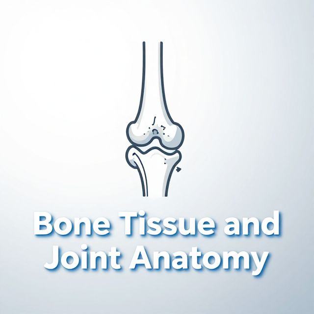
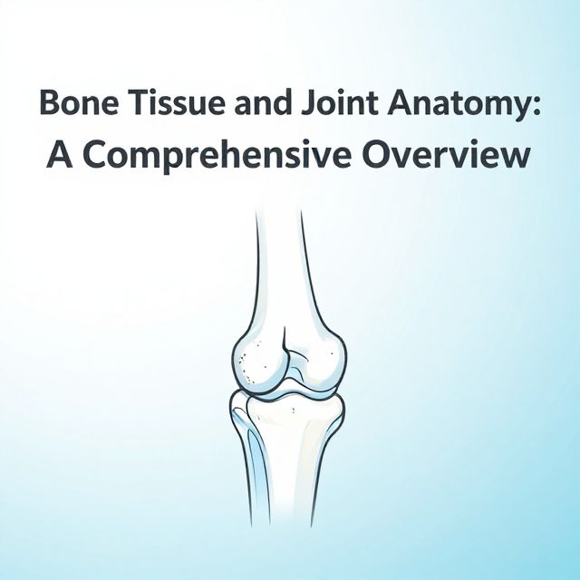
🦴 Bone Tissue and Joint Anatomy: A Comprehensive Study Guide

Bone tissue is a specialized connective tissue vital for structural support, protection, and mineral homeostasis. Its unique properties stem from a complex blend of inorganic and organic components, contributing to both rigidity and flexibility. Understanding its composition, structure, cellular elements, formation, degradation, and associated joints is crucial for comprehending the musculoskeletal system.

1. Bone Composition 📚

Bone tissue is a composite material, primarily made of:

  • Inorganic Component (approx. 65%)
    • Calcium phosphate: Forms pin-shaped hydroxyapatite crystals, providing hardness and rigidity.
    • Water
    • Other minerals: Bicarbonate, citrate, magnesium, sodium, potassium.
  • Organic Component (approx. 35%)
    • Fibers & Ground Substance:
      • Type I Collagen: Accounts for 95% of the organic component, providing flexibility and tensile strength.
      • Ground Substance: Composed of Glycosaminoglycans (GAGs) and Proteoglycans.
    • Glycoproteins: Osteonectin, osteocalcin, osteopontin, bone sialoprotein (involved in mineralization and cell adhesion).

2. Bone Structure: Types of Secondary Bone Tissue 📊

Bone tissue is broadly classified into two structural types:

2.1. Spongy Bone (Cancellous Bone)

  • Location: Found in the metaphysis and epiphysis of short and long bones, and in squamous bones.
  • Organization: Highly organized into bone trabeculae (spiculae).
  • Structure: Contains parallel lamellae.
  • Marrow: Bone marrow is located between the trabeculae.
  • Nutrient Acquisition: Cells receive nutrients from vessels within the bone marrow via cytoplasmic extensions.

2.2. Compact Bone (Cortical Bone)

  • Location: Forms the external aspects of all bones, providing strength and protection.
  • Structure: Contains canals, osteons (Haversian systems), and other lamellar structures.

Canals of Compact Bone

  • Haversian Canals: Run parallel to the longitudinal axis of the bone.
  • Volkmann Canals: Connect Haversian canals, running perpendicularly.
  • Vessel Connection: Vessels reach Haversian canals by passing through Volkmann canals. These vessels are surrounded by loose connective tissue containing nerve fibers.

Types of Lamellae

  • Special Lamellae: Form the concentric rings of osteons (Haversian systems).
  • Circumferential Lamellae:
    • Inner Circumferential: Line the inner surface of the compact bone.
    • Outer Circumferential: Line the outer surface, just beneath the periosteum.
  • Interstitial Lamellae: Irregular fragments of old osteons located between intact osteons and circumferential lamellae.

3. Bone Cells 🔬

Five main cell types are responsible for bone formation, maintenance, and degradation:

3.1. Osteoprogenitor (Osteogenic) Cells

  • Origin: Mesenchymal cells committed to becoming bone cells.
  • Morphology: Fusiform, similar to fibroblasts.
  • Activity: Divide by mitosis and proliferate.
  • Differentiation:
    • Differentiate into osteoblasts under physiological conditions (e.g., osteon degeneration-regeneration, bone fracture).
    • Can differentiate into chondrogenic cells at low oxygen levels.
  • Organelles: Few GER, underdeveloped Golgi complex, abundant free ribosomes.
  • Localization: Inner aspect of periosteum, connective tissue of Haversian & Volkmann canals, endosteum.

3.2. Bone-Lining Cells

  • Nature: An intermediate cell type between osteoprogenitor cells and osteoblasts.
  • Properties: Similar to osteoprogenitor cells but cannot divide.
  • Function: Quiescent cells residing on the bone surface. They activate upon appropriate signals to produce matrix.
  • Connections: Have gap junctions (nexus) between them.

3.3. Osteoblasts

  • Differentiation: From osteoprogenitor cells, influenced by BMP (Bone Morphogenetic Protein) and TGF-β (Transforming Growth Factor-beta).
  • Arrangement: Align in a single row, polarized.
  • Connections: Have gap junctions (nexus) with each other and with osteocytes.
  • Morphology: Cuboidal or oval-shaped, depending on activity state.
  • Organelles: Well-developed GER and Golgi complex, abundant secretory granules (e.g., alkaline phosphatase, pyrophosphatase). Euchromatic nucleus.
  • Products:
    • Type I collagen
    • Osteocalcin, Osteonectin (involved in mineralization)
    • Osteopontin (forms sealing zone for osteoclasts)
    • Sialoprotein (binder to extracellular matrix)
    • Osteoprotegerin, M-CSF (macrophage-colony stimulating factor), RANKL (ligand for receptor activation of nuclear factor kappa B)
    • Receptor for parathyroid hormone
    • Osteoclast-stimulating factor

3.4. Osteocytes

  • Differentiation: Osteoblasts become entrapped within the uncalcified bone matrix (osteoid) they produce. As the tissue calcifies, osteoblast activity decreases, and they transform into squamous-shaped osteocytes.
  • Location: Reside within oval/squamous-shaped lacunae. Randomly oriented in primary bone, between lamellae in secondary bone.
  • Structure: Cytoplasmic extensions extend through canaliculi, connecting with each other via gap junctions.
  • Organelles: Heterochromatic nucleus, poor in organelles, decreased GER and Golgi compared to osteoblasts.
  • Nutrition: Provided via cytoplasmic extensions.
  • Aging: Cytoplasmic extensions shorten, and connections are lost, leading to matrix degeneration if osteocytes die.

3.5. Osteoclasts

  • Function: Degrade bone tissue, controlling blood calcium levels.
  • Origin: Monocytes fuse to form multinuclear osteoclasts.
  • Location: Reside at the surface of trabeculae or the inner aspect of compact bone, specifically within Howship Lacunae.
  • Cytoplasm: Acidophilic.
  • Organelles: Cytoplasmic extensions at the bone tissue aspect, rich in mitochondria, well-developed Golgi, lysosomal enzymes (collagenase, acid phosphatase).

Howship Lacunae 💡

This is the resorption pit where osteoclasts degrade bone. It has distinct zones:

  • Ruffled Border: Area of active degradation.
  • Clear Zone: Immediately neighbors the ruffled border, free of organelles but rich in actin.
  • Vesicular Zone: Contains endo- and exocytotic vesicles.
  • Basal Zone: Farthest from the lacuna, contains organelles and nuclei.
  • Sealing Zone: Osteopontin from bone and actin from osteoclast bind via integrins, creating a sealed environment for degradation.

4. Bone Degradation by Osteoclasts 📉

1️⃣ Osteoclasts contain carbonic anhydrase, which converts CO2 and H2O into carbonic acid. 2️⃣ Carbonic acid dissociates into hydrogen (H+) and bicarbonate (HCO3-) ions. 3️⃣ The osteoclast uses a proton pump to actively secrete H+ ions into the Howship lacuna, significantly decreasing the pH (acidic environment). 4️⃣ This acidic environment degrades the inorganic component of the bone matrix. 5️⃣ Lysosomal enzymes (e.g., collagenase) then degrade the organic components. 6️⃣ Tissue fragments are endocytosed, further degraded, and then expelled into the nearest capillary.

5. Bone Sheaths 🛡️

Bone is covered by two protective sheaths:

5.1. Periosteum

  • Location: Outer surface of bone.
  • Layers:
    • Outer Fibrous Layer: Dense irregular connective tissue.
    • Inner Cellular Layer: Contains osteoprogenitor cells.
  • Sharpey Fibers: Collagen fibers originating from the outer fibrous layer that extend into the outer circumferential and outer interstitial lamellae. Abundant at tendon/ligament connections to bone.

5.2. Endosteum

  • Location: Lines the inner aspect of compact bone, Haversian canals, and surfaces of spongy bone trabeculae.
  • Composition: Reticular connective tissue.
  • Cells: Contains osteoprogenitor cells.

6. Ossification (Bone Formation) 🏗️

Bone formation occurs through two main processes, both involving initial bone degradation to achieve the final shape. Bone development is controlled by hormones like growth hormone, parathyroid hormone, sex steroids, and calcitonin.

6.1. Intramembranous Ossification

  • Process: Direct differentiation of mesenchymal tissue into bone tissue.
  • Steps:
    1. Mesenchymal cells divide, differentiate into osteoprogenitor cells, then into osteoblasts.
    2. Osteoblasts produce bone matrix (osteoid).
    3. Capillaries supply calcium and phosphorus, leading to osteoid calcification.
    4. Primary bone is formed, then organized into secondary bone tissue.
  • Development: Forms compact parts of short and long bones.
  • Bony Collar: Initially formed at the diaphysis periphery, it inhibits chondrocyte nourishment, forcing degeneration. Osteoclasts then pierce it to create nutritive pores for vessels and osteoprogenitor cells.
  • Marrow Formation: Migrated mesenchymal and hematopoietic elements differentiate into bone marrow.

6.2. Endochondral Ossification

  • Process: Bone tissue replaces a pre-existing hyaline cartilage model.
  • Development: Forms spongy parts of short and long bones.
  • Steps:
    1. A hyaline cartilage model forms (e.g., at the epiphysis & metaphysis interface).
    2. Primary Ossification Center: Forms in the diaphysis as vessels carry ions, calcifying the matrix.
    3. Secondary Ossification Centers: Appear in the epiphysis as ossification proceeds, with progenitor cells reaching these areas.
    4. Epiphyseal Plate: The cartilage model between primary and secondary ossification centers, responsible for longitudinal growth. It is typically lost around age 20.
  • Growth: Chondrocytes divide, lengthening the cartilage model. The bony collar thickens, blocking nutrition and promoting bone formation.

6.3. Thickening of Bone

  • Mechanism: Occurs via appositional growth.
  • Process: Osteoprogenitor cells of the periosteum differentiate into osteoblasts, adding new bone tissue to the subperiosteal zone. Simultaneously, the inner aspect of the bone is eroded to enlarge the bone marrow cavity.

7. Bone Health and Diseases ⚠️

Imbalances in bone remodeling can lead to various conditions:

  • Osteoporosis: Loss of bone mass, increased osteoclast number.
  • Osteomalacia: Softening of bone due to defective osteoid mineralization (e.g., Vitamin D insufficiency, renal/intestinal disease).
    • Rickets: Mineralization defect at the epiphyseal plate in children.
  • Osteopetrosis: Dysfunction of osteoclasts, leading to dense, brittle bones.
  • Osteosclerosis: Hyperactivity of osteoblasts, resulting in abnormally dense bone.

Substances Affecting Bone Metabolism 💊

  • Decreasing Bone Degradation: Calcium, Vitamin D, Estrogen, Calcitonin, Biphosphonates (reduce osteoclast activity/number).
  • Increasing Bone Formation: Fluoride, Androgen, Growth Hormone, Statins (stimulate osteoblast differentiation).

8. Joint Types 🤸‍♀️

Joints are critical for movement and stability, classified by their structure and degree of movement:

8.1. Synarthrosis (Immovable or Slightly Movable Joints)

  • Fibrous Joints: Connected by fibrous connective tissue.
    • Examples: Tooth-alveolar (gomphosis), radioulnar (syndesmosis), tibiofibular (syndesmosis).
  • Cartilaginous Joints: Connected by cartilage.
    • Synchondrosis: Hyaline cartilage (e.g., sternocostal joints).
    • Symphysis: Fibrocartilage (e.g., vertebral discs, symphysis pubis).
  • Synostosis: Bones fused by bone tissue (e.g., adult skull sutures).

8.2. Synovial Joint (Diarthrosis - Freely Movable Joints)

  • Joint Capsule: Encloses the joint cavity.
    • Fibrous Capsule: Dense connective tissue, continuous with the periosteum.
    • Synovium: Lines the inner aspect of the fibrous capsule, but does not line the joint cartilage. Rich in vessels and cells.

Synovium

  • Synovial Lining Layer (Intima):
    • Type A Synoviocytes: Macrophage-like cells, involved in degradation ("garbage-degrader cells"). Prominent GER, Golgi, many lysosomes.
    • Type B Synoviocytes: Fibroblast-like cells, synthesize matrix and synovial fluid. Prominent GER, some processes, vacuoles.
    • Type C Synoviocytes: Intermediate type cells.
  • Subsynovial Tissue (Stroma): Beneath the intima.

Synovial Fluid

  • Composition: Water and solutes similar to blood transudate and interstitial tissue fluid.
  • Products of Type B Synoviocytes: Hyaluronic acid, Lubrisin (glycoprotein).
  • Cells: Contains some lymphocytes and monocytes.

Functions of Synovium and Synovial Fluid ✅

  • Decreasing friction between articular cartilages.
  • Nourishment of articular cartilage.
  • Metabolic waste disposal.
  • Providing joint stability.

Kendi çalışma materyalini oluştur

PDF, YouTube videosu veya herhangi bir konuyu dakikalar içinde podcast, özet, flash kart ve quiz'e dönüştür. 1.000.000+ kullanıcı tercih ediyor.

Sıradaki Konular

Tümünü keşfet
Bone Tissue and Joint Anatomy

Bone Tissue and Joint Anatomy

An academic summary detailing the composition, structure, cellular components, development, and pathologies of bone tissue, alongside an overview of joint types and synovial joint anatomy.

14 dk Özet 25 15
Anatomy and Physiology of the Oral Cavity

Anatomy and Physiology of the Oral Cavity

Explore the detailed anatomy and physiology of the oral cavity, including teeth, gingivae, lips, cheeks, palate, and tongue, along with their vascular and neural supplies.

14 dk Özet 25 15
Body Systems for Strength Training

Body Systems for Strength Training

Explore the musculoskeletal and neuromuscular systems essential for human movement and strength, covering bones, joints, muscles, and contraction mechanisms.

8 dk Özet 25 15
Understanding the Respiratory System: Alveoli, Pleura, and Lung Health

Understanding the Respiratory System: Alveoli, Pleura, and Lung Health

Explore the intricate details of the human respiratory system, focusing on alveolar function, pleural membranes, breathing mechanics, and common lung diseases.

5 dk Özet
Mastering Your Upper Limb Practical Exam

Mastering Your Upper Limb Practical Exam

Feeling nervous about your upper limb practical exam? This podcast breaks down the essential anatomy you need to know, from bones and muscles to nerves and vessels, to help you ace it!

Özet 23 10
Basic Anatomy and Physiology of the Autonomic Nervous System

Basic Anatomy and Physiology of the Autonomic Nervous System

This podcast provides a detailed educational overview of the basic anatomy and physiology of the Autonomic Nervous System, its role in homeostasis, and its distinction from the somatic system.

Özet 25
The Musculoskeletal System: Structure, Function, and Locomotion

The Musculoskeletal System: Structure, Function, and Locomotion

Explore the intricate musculoskeletal system, its components, functions, and the mechanisms of locomotion in various organisms, with a detailed focus on the human body.

Özet 25 15 Görsel
The Digestive System: An Academic Overview

The Digestive System: An Academic Overview

An academic summary of the human digestive system, detailing its organs, processes, and functions, from mechanical breakdown to nutrient absorption and waste elimination.

4 dk Özet 25 15