Study Material: The Respiratory System
Source Information: This study material has been compiled from a lecture audio transcript and supplementary copy-pasted text (likely from a PDF or PowerPoint presentation).
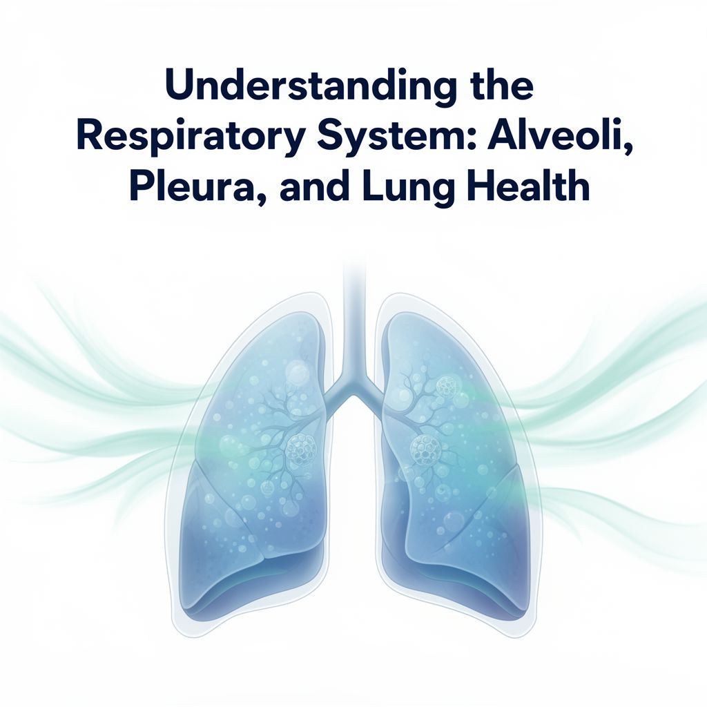
📚 Introduction to the Respiratory System
This guide provides an in-depth look at the fundamental components and functions of the respiratory system. We will explore everything from the smallest structural units of the lungs to their protective membranes, the mechanics of respiratory movements, and significant diseases affecting the lungs. The aim is to help you better understand the complex structure and vital importance of the respiratory system for our body.
🌬️ I. The Alveoli: Gas Exchange and Protection
The alveoli are the primary functional units of the lungs, crucial for gas exchange.
A. Surfactant Film
- Function:
- Lowers surface tension at the air-epithelium interface. ✅
- Helps prevent alveolar collapse during exhalation. ✅
- Allows alveoli to inflate with less inspiratory force, easing the work of breathing. ✅
- Composition & Turnover:
- Produced by Type II alveolar cells.
- Constantly renewed; gradually removed by pinocytosis in both Type I and Type II alveolar cells, and by macrophages.
- Fetal Development:
- Appears in the last weeks of gestation as Type II cells differentiate and form lamellar bodies. 💡
B. Infant Respiratory Distress Syndrome (IRDS)
- Cause:
- Leading cause of death in premature babies. ⚠️
- Due to incomplete differentiation of Type II alveolar cells.
- Results in a deficit of surfactant.
- Causes difficulty in expanding the alveoli during breathing.
- Treatment:
- Insertion of an endotracheal tube. 1️⃣
- Provides continuous positive airway pressure (CPAP). 2️⃣
- Administers exogenous surfactant (chemically synthesized or purified from animal lungs). 3️⃣
C. Alveolar Macrophages (Dust Cells)
- Location: Found in alveoli and the interalveolar septum.
- Function:
- Phagocytose erythrocytes lost from damaged capillaries.
- Clear airborne particulate matter that has penetrated the alveoli.
- Origin: Tens of millions of monocytes migrate daily from the microvasculature into the lung tissue.
D. Alveolar Lining Fluids
- Removal: Removed via the conducting passages through ciliary activity.
- Formation: As secretions move up the airways, they combine with bronchial mucus to form bronchoalveolar fluid.
- Function:
- Helps remove particulate components from inspired air.
- Bacteriostatic: Contains lysozyme and other protective agents.
- Production: Protective agents are produced by bronchiolar exocrine cells, Type II alveolar cells, and alveolar macrophages.
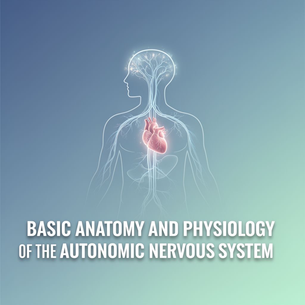
🩸 II. Lung Vasculature and Nerves
The lungs have a dual blood supply and are extensively innervated.
A. Blood Circulation
- Pulmonary Circulation: Carries O2-depleted blood for gas exchange.
- Bronchial Circulation: Carries O2-rich blood to supply lung tissue.
B. Innervation
- Autonomic Fibers: Both parasympathetic and sympathetic fibers innervate the lungs.
- Control: These fibers control reflexes regulating smooth muscle contractions, which determine the diameters of the airways. 📈
🛡️ III. Pleural Membranes
The lungs are encased by protective serous membranes.
A. The Pleura
- Definition: A serous membrane covering the lung's outer surface and the internal wall of the thoracic cavity.
- Layers:
- Visceral Pleura: Attached directly to the lung tissue.
- Parietal Pleura: Lines the thoracic walls.
- Continuity: The two layers are continuous at the hilum (the point where structures like blood vessels and nerves enter the organ).
- Composition: Both layers are composed of simple squamous mesothelial cells on a thin connective tissue layer containing collagen and elastic fibers.
- Elastic Fibers: The elastic fibers of the visceral pleura are continuous with those of the pulmonary parenchyma.
B. Pleural Cavity and Fluid
- Pleural Cavity: The narrow space between the parietal and visceral layers.
- Lining: Entirely lined with mesothelial cells.
- Serous Fluid: Mesothelial cells produce a thin film of serous fluid.
- Function: Acts as a lubricant, facilitating the smooth sliding of one surface over the other during respiratory movements. ✅
💨 IV. Respiratory Movements
Breathing involves coordinated muscle action and elastic recoil.
A. Inhalation (Inspiration)
- Muscle Contraction:
- External intercostal muscles contract, elevating the ribs.
- Diaphragm contracts, lowering the bottom of the thoracic cavity.
- Volume Change: Increases the diameter of the thoracic cavity, leading to pulmonary expansion.
- Airway Changes: Bronchi and bronchioles increase in diameter and length.
- Alveolar Expansion: The respiratory portion enlarges, mainly due to the expansion of alveolar ducts; individual alveoli enlarge only slightly.
- Elastic Fibers: The elastic fibers of the pulmonary parenchyma are stretched by this expansion.
B. Exhalation (Expiration)
- Passive Process: Lungs retract passively.
- Muscle Relaxation: Due to relaxation of the external intercostal muscles and diaphragm.
- Elastic Recoil: Elastic fibers return to their unstretched condition, pushing air out.
🩺 V. Lung Cancer
Lung cancer is a common and serious disease with various forms.
A. General Information
- One of the most common forms of cancer. ⚠️
B. Types of Lung Cancer
- Squamous Cell Carcinoma:
- Closely correlated with a history of smoking. 🚬
- Arises most often from epithelial cells of segmental bronchi.
- Adenocarcinoma:
- The most common lung cancer in nonsmokers.
- Usually arises from epithelial cells more peripherally, in bronchioles and alveoli.
- Small Cell Carcinoma:
- Less common but highly malignant.
- Develops after neoplastic transformation of small granule Kulchitsky cells (a type of endocrine cell) in bronchial respiratory epithelium.