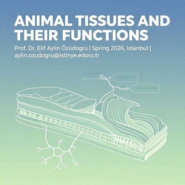
📚 Histology Study Material: Glands and Membranes
Source Information: This study material has been compiled from a copy-pasted text and a lecture audio transcript on Histology, covering the topics of glands and various biological membranes.
Introduction to Glands and Signaling
Glands are specialized epithelial structures responsible for synthesizing and secreting various substances essential for bodily functions. They are broadly categorized based on how they release their products. Beyond the major classifications, cells also employ localized signaling mechanisms.
1. Gland Classification by Product Release
Glands are primarily classified into two major groups based on their secretion pathway:
- Exocrine Glands ✅
- Secrete their products onto a surface (internal or external) directly or through epithelial ducts or tubes that connect to a surface.
- Examples: Sweat glands, salivary glands, sebaceous glands.
- Endocrine Glands ✅
- Secrete their products, known as hormones, into the surrounding connective tissue.
- From the connective tissue, hormones enter the bloodstream to reach distant target cells.
- Key Characteristic: Lack ducts.
- Examples: Thyroid gland, adrenal gland, pituitary gland.
2. Localized Cellular Signaling
Beyond systemic endocrine signaling, cells can communicate locally:
- Paracrine Signaling 💡
- Individual cells secrete substances that do not reach the bloodstream but affect other nearby cells.
- Substances are released into the subjacent extracellular matrix.
- Example: Endothelial cells of blood vessels release factors that impact adjacent vascular smooth muscle cells, causing contraction or relaxation of the vascular wall.
- Autocrine Signaling 💡
- Cells secrete molecules that bind to receptors on the same cell that released them.
- Often initiates negative feedback pathways to modulate their own secretion.
- Example: Cells of the immune system, involving interleukin signaling molecules.
Exocrine Gland Secretion Mechanisms
Exocrine glands utilize three fundamental mechanisms to release their secretory products:
- Merocrine Secretion ✅
- Mechanism: The secretory product is delivered in membrane-bounded vesicles to the apical surface of the cell. These vesicles fuse with the plasma membrane and extrude their contents by exocytosis.
- Prevalence: This is the most common mechanism of secretion.
- Example: Pancreatic acinar cells.
- Apocrine Secretion ✅
- Mechanism: The secretory product is released in the apical portion of the cell, surrounded by a thin layer of cytoplasm within an envelope of plasma membrane. A portion of the cell's cytoplasm is lost with the secretion.
- Example: Found in the lactating mammary gland, responsible for releasing large lipid droplets into the milk.
- Holocrine Secretion ✅
- Mechanism: The secretory product accumulates within the maturing cell, which simultaneously undergoes destruction orchestrated by programmed cell death (apoptosis). Both secretory products and cell debris are discharged into the lumen of the gland. The entire cell is lost during secretion.
- Example: Sebaceous glands of the skin and tarsal (Meibomian) glands of the eyelid.
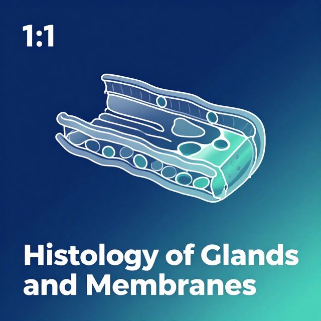
Exocrine Gland Classification by Structure
Exocrine glands are classified as either unicellular or multicellular.
1. Unicellular Glands
- Structure: Simplest form, consisting of single secretory cells distributed among other non-secretory cells.
- Key Example: Goblet Cell ⚠️ (Important cell!)
- A mucus-secreting cell (stains PAS+ due to mucin content).
- Positioned among other columnar cells (e.g., in the intestines and respiratory tract).
- Polarity: Highly polarized with the nucleus and other organelles concentrated at the base of the cell.
- Apical Region: Contains secretory granules with mucin. The apical plasma membrane projects short microvilli, and the apical portion is shaped like a cup, distended by abundant mucus-laden granules.
- Basal Region: Lacks these granules and is shaped like a stem.
2. Multicellular Glands
- Structure: Composed of more than one cell, exhibiting varying degrees of complexity.
- Simplest Form: A cellular sheet where each surface cell is a secretory cell (e.g., the lining of the stomach and its gastric pits, which secrete mucus).
- Complex Forms: Typically form tubular invaginations from the surface.
- End Pieces: Contain the secretory cells.
- Duct: Connects the secretory cells to the surface.
Subclassification of Multicellular Glands:
- Based on Duct Branching:
- Simple Gland: If the duct is unbranched.
- Compound Gland: If the duct is branched.
- Based on Secretory Portion Shape:
- Tubular: Secretory portion is shaped like a tube (can be straight, branched, or coiled).
- Alveolar (or Acinar): Secretory portion is shaped like a flask or grape (can be single or branched).
- Tubuloalveolar: Secretory portion is a tube ending in a sac-like dilation.
Gland Classification by Secretion Type
Glands are also named based on the type of secretion they produce:
- Mucous Secretions 📚
- Characteristics: Viscous and slimy.
- Examples: Goblet cells, secretory cells of the sublingual salivary glands, surface cells of the stomach.
- Biochemistry: Result from extensive glycosylation of constituent proteins with anionic oligosaccharides.
- Histology: Mucinogen granules are PAS positive. They are water-soluble and often lost during routine tissue preparation, making the cytoplasm of mucous cells appear empty in H&E stained paraffin sections.
- Nucleus: Usually flattened against the base of the cell by accumulated secretory product.
- Serous Secretions 📚
- Characteristics: Watery, poorly glycosylated or non-glycosylated protein secretions.
- Examples: Parotid gland, pancreas.
- Histology: Nucleus is typically round or oval. Apical cytoplasm is often intensely stained with eosin if secretory granules are well preserved. Perinuclear cytoplasm often appears basophilic due to extensive rough endoplasmic reticulum (RER), characteristic of protein-synthesizing cells.
- Mixed Glands
- Some glands contain both mucous and serous cells (e.g., submandibular gland).
- In routine tissue preparation, serous cells are often located further from the lumen of the acinus and appear as crescents or demilunes (half-moons) at the periphery of mucous acini.
Biological Membranes
In specific locations, surface epithelium and its underlying connective tissue form a functional unit called a membrane. There are four main types of biological membranes:
- Cutaneous Membranes (Skin) 🌍
- Exposed directly to the air.
- Consists of epidermis (stratified squamous epithelium) and dermis (connective tissue).
- Mucous Membranes (Mucosa) 🌊
- Location: Wet membranes that open to the exterior (e.g., respiratory, urinary, digestive tracts).
- Components:
- Surface epithelium (with or without glands).
- Supporting connective tissue called the lamina propria.
- A basement membrane separating the epithelium from the lamina propria.
- Sometimes a layer of smooth muscle called the muscularis mucosae as the deepest layer.
- Serous Membranes (Serosa) 🛡️
- Location: Line body cavities that are completely separate from the exterior (e.g., peritoneal, pericardial, and pleural cavities).
- Exception: In females, the peritoneal cavity communicates with the exterior via the genital tract.
- Components:
- A lining epithelium called the mesothelium.
- A supporting connective tissue.
- A basement membrane between the two.
- Characteristics: Do not contain glands, but the fluid on their surface is watery.
- Layers:
- Parietal serosa: Lines internal body cavity walls.
- Visceral serosa: Covers internal organs (viscera).
- These double layers are separated by a slit-like cavity filled with serous fluid, secreted by both layers.
- Location: Line body cavities that are completely separate from the exterior (e.g., peritoneal, pericardial, and pleural cavities).
- Synovial Membranes 🦵
- Location: Found at joints (e.g., knee, shoulder).
- Structure: Consist of areolar tissue with an incomplete layer of overlying epithelium.
Practice Questions
-
Which of the following is NOT true about the cell which is represented by A in the micrograph (referring to a goblet cell)? a) It is a unicellular exocrine gland. b) It is positioned among the epithelial cells of the urinary tract. c) It contains secretory granules at the apical part. d) It is a highly polarized cell. e) It is PAS (+). Answer: b) (Goblet cells are typically found in the intestines and respiratory tract, not primarily the urinary tract).
-
Which of the following is NOT true about the gland tissue/cell? a) Mucous cells produce viscous and slimy secretion. Answer: (The question is incomplete, but based on the provided options, 'a' is a true statement about mucous cells.)