Understanding Connective Tissues and Their Cells - kapak
Bilim#connective tissue#histology#cells#extracellular matrix

Understanding Connective Tissues and Their Cells

Explore the fundamental types, structures, and cellular components of connective tissues, including their crucial roles in the body's support, protection, and immunity.

rana_lApril 11, 2026 ~16 dk toplam
01

Flash Kartlar

25 kart

Karta tıklayarak çevir. ← → ile gez, ⎵ ile çevir.

1 / 25
Tüm kartları metin olarak gör
  1. 1. What is the defining characteristic of all connective tissues?

    The defining characteristic of all connective tissues is the extracellular matrix. This matrix is actively produced by the connective tissue cells themselves and varies significantly in composition depending on the specific tissue type. It is composed of fibers like collagen and elastin, and a ground substance.

  2. 2. From which embryonic cells do connective tissues originate?

    Connective tissues originate from mesenchymal cells. These are embryonic cells that possess the capability to differentiate into various specialized forms, including those found in connective tissue proper, cartilage, bone, and even hematopoietic and endothelial cells, highlighting their versatility in tissue formation.

  3. 3. List three crucial roles of the extracellular matrix in connective tissues.

    The extracellular matrix plays several crucial roles. It enables organ mobility, allowing organs to move relative to each other. It transmits forces, providing mechanical strength and support. Additionally, it offers stability as a firm construction and facilitates the diffusion of molecules among cells, which is vital for nutrient and waste exchange.

  4. 4. How are connective tissue proper types broadly classified, and what distinguishes them?

    Connective tissue proper is broadly classified into loose and dense types. Loose connective tissue is characterized by a good blood supply, innervation, and immune cells, allowing for mobility and shape changes. Dense connective tissue, conversely, is dominated by extracellular matrix fibers, providing strength and resistance to stress.

  5. 5. Describe the key features of loose connective tissue.

    Loose connective tissue is characterized by a good blood supply, innervation, and the presence of immune cells. It allows for mutual mobility and shape changes in various membranes, filling gaps between organs. This tissue type is crucial for flexibility and supporting other tissues and organs.

  6. 6. What is the primary function of adipose connective tissue?

    Adipose connective tissue, a type of loose connective tissue, is vital for metabolism, hormone production, and energy storage. It stores neutral lipids as triglycerides, acting as a significant energy reserve. Its rich blood supply supports these metabolic and endocrine functions.

  7. 7. Differentiate between regular and irregular dense connective tissue, providing an example for each.

    Regular dense connective tissue has fibers running in one direction, providing strength in a single plane, such as in tendons and ligaments. Irregular dense connective tissue has fibers running in various directions, offering strength and resistance to stress from multiple directions, as seen in the corium of the skin or organ capsules.

  8. 8. Name three types of connective tissue based on their predominant fiber type.

    Connective tissues can be categorized by their predominant fiber type into collagenous, elastic, or reticular tissues. Collagenous tissues are rich in collagen fibers, providing tensile strength. Elastic tissues contain abundant elastin, allowing for flexibility and recoil. Reticular tissues, with fine reticular fibers, form delicate supporting networks.

  9. 9. What are the main components that form the composite structure of cartilage?

    Cartilage features a composite structure primarily made of cartilage cells, known as chondrocytes, which are embedded within an extracellular matrix. This matrix is rich in hydrophilic molecules, giving cartilage its unique properties.

  10. 10. Explain how the extracellular matrix of cartilage contributes to its function.

    The extracellular matrix of cartilage is rich in hydrophilic molecules, which enables significant resistance to compression. This composition provides cartilage with a semi-rigid consistency, allowing it to act as a shock absorber and provide structural support while maintaining some flexibility.

  11. 11. Which type of cartilage is the most common, and where can it be found in the body?

    Hyaline cartilage is the most common type of cartilage. It is found in the fetal skeleton, serving as a template for bone formation, in joint cartilages where it reduces friction, and reinforcing structures like the trachea and bronchi, providing structural support to airways.

  12. 12. What distinguishes elastic cartilage from other types, and where is it located?

    Elastic cartilage is distinguished by its richness in elastin fibers, which provides flexible support. This allows it to return to its original shape after deformation. It is typically found in structures requiring significant flexibility, such as the epiglottis and the auricle (external ear).

  13. 13. Describe the characteristics and primary location of fibrous cartilage.

    Fibrous cartilage resembles dense irregular collagenous tissue due to its abundance of collagen fibers, making it incredibly strong and resistant to tension. It forms structures that require high tensile strength and shock absorption, such as intervertebral discs and menisci in joints.

  14. 14. Why does mature cartilage have limited healing ability?

    Mature cartilage has a very low metabolism and is avascular, meaning it lacks a direct blood supply. It relies on diffusion for nutrition from surrounding tissues. This limited nutrient supply and metabolic activity contribute significantly to its poor regenerative capacity and limited healing ability after injury.

  15. 15. What are the two broad categories of cells found in connective tissues?

    The two broad categories of cells found in connective tissues are resident cells and wandering or transient cells. Resident cells are permanent inhabitants of the tissue, while wandering cells migrate into the tissue from the circulatory system, often in response to specific stimuli.

  16. 16. Which cell is the most common resident cell of connective tissue proper, and what is its primary function?

    The fibroblast is the most common resident cell of connective tissue proper. It is metabolically and synthetically active, primarily responsible for producing both the fibrous components (like collagen and elastin) and the amorphous ground substance of the extracellular matrix.

  17. 17. What is the resting form of a fibroblast called, and what is its role?

    The resting form of a fibroblast is called a fibrocyte. Fibrocytes are less metabolically active than fibroblasts and stop dividing. Their primary role is to maintain the existing extracellular matrix and form a supporting structure for the tissue, essentially acting as maintenance cells.

  18. 18. What are adipocytes specialized for, and what do they store?

    Adipocytes, or fat cells, are specialized for the uptake, collection, metabolism, and release of neutral lipids. They store these lipids primarily as triglycerides, serving as the body's main energy reserve. They also act as a source and target for various hormones.

  19. 19. Differentiate between unilocular and multilocular adipocytes.

    Unilocular adipocytes store a single, large lipid droplet, giving them a "signet ring" appearance, and are primarily involved in energy storage. Multilocular adipocytes contain multiple smaller lipid droplets and are specialized for heat production through non-shivering thermogenesis, often found in newborns and hibernating animals.

  20. 20. What is the origin of macrophages, and what are their key functions?

    Macrophages originate from monocytes, which are white blood cells that migrate from the circulatory system into tissues. They are highly motile and capable of phagocytosis, engulfing cellular debris, pathogens, and foreign substances. They also play a crucial role in presenting antigens to other leukocytes, initiating immune responses.

  21. 21. What substances are contained within the large basophilic granules of mastocytes, and when are they released?

    Mastocytes (mast cells) are characterized by large basophilic granules containing substances such as heparin, histamine, and proteases. These substances are released during inflammatory and allergic reactions, causing vasodilation (widening of blood vessels) and increased vascular permeability, contributing to the symptoms of inflammation.

  22. 22. What is the primary function of plasma cells in connective tissue?

    Plasma cells are activated B-lymphocytes. Their primary function is to produce and secrete antibodies. These protein antibodies exhibit sensitive and specific reactivity against particular antigens, playing a critical role in humoral immunity by neutralizing pathogens and toxins.

  23. 23. How does the composition of the extracellular matrix vary among different connective tissue types?

    The composition of the extracellular matrix varies significantly depending on the specific tissue type. This variation is primarily in the types and proportions of fibers (like collagen, elastin, reticular fibers) and the ground substance components, which dictates the tissue's mechanical properties and functions, from flexible support to rigid strength.

  24. 24. What is the significance of mesenchymal cells in the development of connective tissues?

    Mesenchymal cells are embryonic cells that are crucial because they are multipotent, meaning they can differentiate into various specialized cell types. This capability allows them to form all types of connective tissues, including bone, cartilage, and connective tissue proper, making them fundamental to the body's structural development.

  25. 25. Provide an example of how loose connective tissue facilitates organ function.

    Loose connective tissue facilitates organ function by allowing for mutual mobility and shape changes in various membranes, and by filling gaps between organs. For instance, it allows the skin to move freely over underlying muscles and provides a flexible packing material that supports organs while permitting their movement.

02

Bilgini Test Et

15 soru

Çoktan seçmeli sorularla öğrendiklerini ölç. Cevap + açıklama.

Soru 1 / 15Skor: 0

What is the defining characteristic that distinguishes all connective tissues from other tissue types?

03

Detaylı Özet

6 dk okuma

Tüm konuyu derinlemesine, başlık başlık.

📚 Connective Tissues: Structure, Types, and Cells

This study material has been compiled and organized from a lecture audio transcript and a copy-pasted text document, providing a comprehensive overview of connective tissues.


1. Introduction to Connective Tissues 🌍

Connective tissues are fundamental components of the body, providing structural support, connecting other tissues, and facilitating various physiological processes. They are characterized by their diverse cellular and structural properties.

1.1. Defining Characteristics ✅

  • Diverse Cellular and Structural Properties: Connective tissues exhibit a wide range of forms and functions.
  • Extracellular Matrix (ECM): The defining feature of all connective tissues. It is actively produced by the connective tissue cells themselves.
    • Composition: Consists of fibers (e.g., collagen, elastin) and a ground substance (amorphous matrix).
    • Variation: The composition of the ECM varies significantly depending on the specific tissue type, influencing its mechanical properties.
  • Origin: All connective tissues originate from mesenchymal cells, which are embryonic cells capable of differentiating into various specialized forms.

1.2. Functions of the Extracellular Matrix 📊

The ECM plays crucial roles in the body:

  • Organ Mobility: Allows organs to move relative to each other.
  • Force Transmission: Transfers mechanical forces throughout the tissue.
  • Stability: Provides a firm construction, offering stability and support.
  • Diffusion: Facilitates the diffusion of molecules and nutrients among cells.

2. Types of Connective Tissues 🔬

Connective tissues are broadly categorized into several types, each with specialized functions.

2.1. Connective Tissue Proper 🩹

This category includes soft connective tissues, further classified based on the density and arrangement of their fibers.

2.1.1. Loose Connective Tissue

  • Characteristics:
    • Good blood supply and innervation.
    • Presence of immune cells.
    • Allows for mutual mobility and shape changes in membranes.
    • Fills gaps between layers of solid and tubular organs.
  • Example: Adipose Connective Tissue 💡
    • A type of loose connective tissue where adipocytes (fat cells) are dominant.
    • Vital for metabolism, hormone production, and energy storage due to its rich blood supply.
    • Also includes embryonic tissues like mesenchyme and umbilical cord tissue.

2.1.2. Dense Connective Tissue

  • Characteristics: Dominated by extracellular matrix fibers.
  • Classification by Fiber Arrangement:
    • Regular Dense Connective Tissue: Fibers run predominantly in one direction.
      • Examples: Tendons (connect muscle to bone) and ligaments (connect bone to bone).
    • Irregular Dense Connective Tissue: Fibers run in various directions.
      • Examples: Corium of the skin, connective tissue capsules of organs.
  • Classification by Predominant Fiber Type:
    • Collagenous Tissue: Most common (e.g., skin, tendons, ligaments).
    • Elastic Tissue: Contains abundant elastic fibers (e.g., some ligaments).
    • Reticular Tissue: Forms a delicate network (e.g., stroma for white blood cells in bone marrow, lymph nodes, spleen).
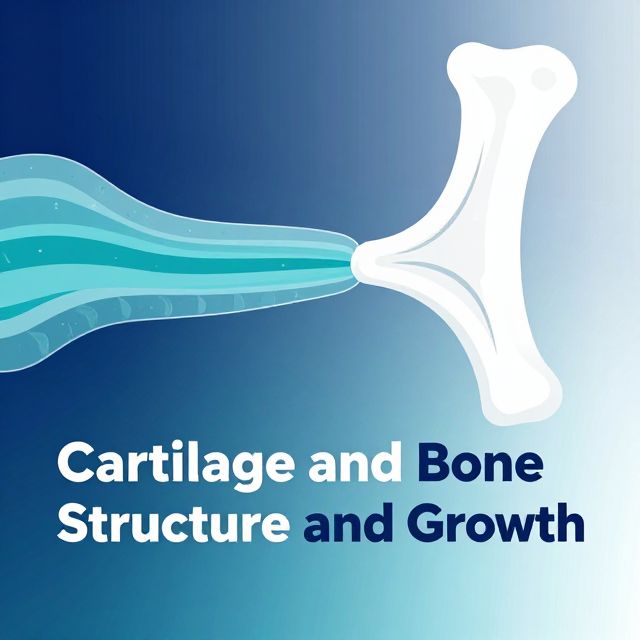
2.2. Cartilage 🦴

Cartilage is a semi-rigid connective tissue characterized by its unique extracellular matrix.

  • General Structure:
    • Chondrocytes: Cartilage cells, occurring individually or in groups.
    • Extracellular Matrix: Fibrous part connected to chondrocytes, rich in high-molecular protein-polysaccharide complexes (ground substance).
    • Hydration: High concentration of hydrophilic molecules ensures hydration, leading to significant resistance to compression and a semi-rigid consistency.
  • Types of Cartilage:
    • Hyaline Cartilage: Most common type.
      • Location: Forms most of the fetal skeleton, joint cartilages, costal cartilages, reinforces trachea and bronchi.
      • Collagen Type: Predominantly Type II collagen.
    • Elastic Cartilage: Elastic due to the presence of elastin.
      • Location: Epiglottis, auricle (earlobe support).
      • Collagen Type: Predominantly Type II collagen.
    • Fibrous Cartilage (Fibrocartilage): Resembles dense irregular collagenous tissue.
      • Characteristics: Very strong due to thick bundles of Type I collagen fibers. Contains chondrocytes and cartilaginous matrix.
      • Location: Outer layer of intervertebral discs, joint discs, menisci.
  • Metabolism & Healing: Mature cartilage has a very low metabolism, is avascular (lacks blood vessels), and relies on diffusion for nutrition (from perichondrium or synovial fluid). This leads to limited healing ability, which further decreases with age.

2.3. Bone Tissue 💀

Bone is a highly specialized, mineralized connective tissue.

  • Composition:
    • Organic Part: Collagen (Type I), glycoproteins, proteoglycans.
    • Mineral Part: Hydroxyapatite crystals.
  • Cells:
    • Osteoblasts: Bone-forming cells, produce the organic matrix.
    • Osteocytes: Mature bone cells, develop from osteoblasts, embedded in lacunae within the mineralized matrix.
    • Osteoclasts: Large, multinucleated cells responsible for bone resorption.
  • Remodeling: Bone tissue continuously undergoes remodeling, a balance between formation (osteoblasts) and resorption (osteoclasts), adjusting to mechanical stress. This process is crucial for healing fractures.
  • Types:
    • Primary Woven Bone: Characterized by random collagen fiber arrangement; replaced during development.
    • Secondary Lamellar Bone: Organized into lamellae (layers) with osteocytes.
      • Osteons (Haversian Systems): Concentric lamellar systems.
    • Compact Bone: Dense outer layer.
    • Spongy (Cancellous/Trabecular) Bone: Inner, porous bone with trabeculae, spaces filled with bone marrow.

3. Key Connective Tissue Cells 🧬

Connective tissues contain a variety of cells, categorized as resident (permanent) or wandering (transient).

3.1. Resident Cells 🏡

These cells are a constant part of the connective tissue and originate locally.

  • 1️⃣ Fibroblast:
    • Function: Most common cell in connective tissue proper. Metabolically and synthetically active, producing fibrous and amorphous intercellular matter.
    • Appearance: Spindle- to star-shaped with multiple protrusions.
    • Cytoplasm: Basophilic due to well-developed granular endoplasmic reticulum (GER) and Golgi apparatus.
  • 2️⃣ Fibrocyte:
    • Function: The resting form of a fibroblast. Stops dividing and forms a supporting structure for the tissue.
    • Appearance: Elongated, spindle-shaped nucleus, predominantly heterochromatin.
  • 3️⃣ Myofibroblast:
    • Function: A variant of fibroblast with smooth muscle cell properties. Contains alpha-smooth muscle actin, allowing contraction. Transmits mechanical forces.
    • Clinical Relevance: Important in wound healing, contributing to wound contraction. Excessive activity can lead to hypertrophic scars or keloids.
  • 4️⃣ Reticular Cell:
    • Function: Specialized fibroblast producing reticular (argyrophilic) fibers, forming a supporting net for lymphocytes and macrophages in lymphoid organs.
    • Appearance: Star-shaped with cytoskeleton-reinforced protrusions.
  • 5️⃣ Adipocyte (Fat Cell) 🍎
    • Function: Specialized for uptake, collection, metabolism, and release of neutral lipids (triglycerides). Source and target for hormones.
    • Origin: Differentiate from adipoblasts (lipoblasts).
    • Types:
      • Unilocular (White) Adipocyte: Contains a single, large lipid droplet, pushing the nucleus and organelles to the periphery (signet-ring appearance). Main component of white adipose tissue.
      • Multilocular (Brown) Adipocyte: Contains multiple, smaller lipid droplets. Nucleus is irregularly positioned. Rich in mitochondria with thermogenin, specialized for heat production (non-shivering thermogenesis). Present in brown adipose tissue.

3.2. Wandering (Transient) Cells 🚶‍♀️

These cells migrate to the connective tissue from the circulatory system, often in response to signals.

  • 1️⃣ Macrophage 🛡️
    • Origin: Differentiate from monocytes.
    • Function: Highly motile, capable of phagocytosis (engulfing damaged cells, pathogens, foreign particles). Present antigens to other leukocytes, initiating immune responses. Release pro-inflammatory mediators and enzymes for tissue remodeling.
    • Appearance: Large, irregular shape with numerous protrusions. Kidney-shaped nucleus.
  • 2️⃣ Mastocyte / Mast Cell (Heparinocyte) 🚨
    • Function: Plays a key role in inflammatory and allergic reactions.
    • Characteristics: Large, ovoid cells with a spherical nucleus. Cytoplasm filled with large, intensely basophilic granules.
    • Granule Content:
      • Heparin: Sulfated glycosaminoglycan, acts as an anticoagulant.
      • Histamine: Derivative of histidine, causes vasodilation, increases endothelial permeability, and supports smooth muscle contraction.
      • Proteases: Serine proteases, metalloproteinases, involved in inflammatory processes.
    • Mechanism: Cell membrane has affinity for IgE antibodies. Binding of antigens to IgE triggers degranulation, releasing granule contents into the extracellular space.
  • 3️⃣ Plasma Cell (Plazmocyte) 💉
    • Origin: Activated B-lymphocytes.
    • Function: Produce and secrete large quantities of specific antibodies (immunoglobulins).
    • Appearance: Elongated, ovoid, or pyramidal shape. Eccentric, round nucleus with a "clock face" or "cart-wheel" chromatin pattern. Large volume of cytoplasm with rich granular endoplasmic reticulum and a prominent Golgi complex.
  • 4️⃣ Pigment Cell (Melanocyte)
    • Origin: Develops from neuroectodermal neural crest cells.
    • Function: Produces, collects, and releases melanin pigments (e.g., eumelanin for black/brown color).
    • Location: Found in epidermis, eye uvea, brain meninges, and connective tissue proper.
    • Appearance: Asymmetrical in epithelium, star-shaped in connective tissue.

Kendi çalışma materyalini oluştur

PDF, YouTube videosu veya herhangi bir konuyu dakikalar içinde podcast, özet, flash kart ve quiz'e dönüştür. 1.000.000+ kullanıcı tercih ediyor.

Sıradaki Konular

Tümünü keşfet
Cartilage and Bone Structure and Growth

Cartilage and Bone Structure and Growth

This summary provides an academic overview of cartilage structure, extracellular matrix, chondrogenesis, distinct cartilage types, and the mechanisms of bone growth and thickening.

6 dk Özet 25 15
Connective Tissue Cells: Structure, Function, and Roles

Connective Tissue Cells: Structure, Function, and Roles

This summary provides an academic overview of connective tissue cells, detailing their classification, morphology, specific functions, and critical roles in tissue maintenance, repair, and immune responses.

9 dk Özet 25 15
Connective Tissue: Structure, Types, and Functions

Connective Tissue: Structure, Types, and Functions

An academic overview of connective tissue, detailing its embryonic origins, classifications into proper and specialized types, and the composition and roles of its fibrous and extracellular matrix components.

7 dk Özet 25 15
Animal Tissues and Their Functions in Vertebrates

Animal Tissues and Their Functions in Vertebrates

An educational overview of the four primary animal tissues in vertebrates: epithelial, connective, muscle, and nervous tissues, including their structure, functions, and locations.

Özet 25 15
Introduction to Basic Genetics

Introduction to Basic Genetics

Explore the fundamental principles of genetics, from cell theory and the nature of genetic material to heredity, cell division, and the diverse branches of genetic study.

Özet 25 15
Understanding Animal Tissues: Structure, Function, and Development

Understanding Animal Tissues: Structure, Function, and Development

Explore the intricate world of animal tissues, focusing on epithelium and connective tissues. Learn about their embryonic origins, diverse structures, and vital functions in the body.

8 dk Özet 25 15
The Musculoskeletal System: Structure, Function, and Locomotion

The Musculoskeletal System: Structure, Function, and Locomotion

Explore the intricate musculoskeletal system, its components, functions, and the mechanisms of locomotion in various organisms, with a detailed focus on the human body.

Özet 25 15 Görsel
Histology of Glands and Membranes

Histology of Glands and Membranes

An academic overview of glandular classification, secretion mechanisms, and the structural and functional characteristics of various biological membranes in histology.

5 dk Özet 25 15