This study material is compiled from a detailed anatomical text and a corresponding lecture audio transcript.
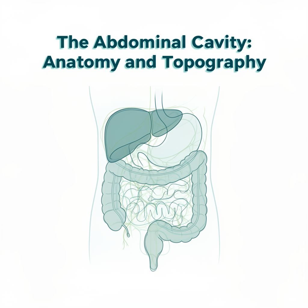
Abdominal Cavity: An Anatomical Overview
This study guide provides a comprehensive overview of the abdominal cavity, its peritoneal coverings, topographical organization, and key neurovascular and lymphatic structures. It aims to clarify the complex anatomy of this vital region of the human body.
1. The Abdominal Cavity and Peritoneum
1.1. Abdominal Cavity Overview
The abdominal cavity is a crucial part of the larger abdominopelvic cavity, which constitutes the sub-diaphragmatic portion of the trunk. ✅ Boundary: The conventional plane passing through the promontory and the superior extremity of the pubic symphysis defines the limit between the abdominal and pelvic cavities, which communicate with each other.
1.2. The Peritoneum (Tunica Serosa) 📚
The peritoneum is a serous membrane originating from the mesothelial cells of embryonic somatopleura and splanchnopleura. It separates from the pleura and pericardium during the 6th week of gestation.
1.2.1. Peritoneal Layers
The peritoneum consists of two continuous layers:
- Visceral Peritoneum:
- Thin and adherent.
- Invests intra-peritoneal organs, anchoring them to the abdominal wall and linking them together.
- Parietal Peritoneum:
- Thicker and less adherent than the visceral peritoneum.
- Lines the abdominopelvic cavity walls and partially covers extra-peritoneal organs.
1.2.2. Peritoneal Spaces
Spaces formed between the parietal peritoneum and the abdominopelvic cavity walls:
- Supra-peritoneal space
- Pre-peritoneal space
- Right lateral peritoneal space
- Left lateral peritoneal space
- Retro-peritoneal space
- Sub-peritoneal space
1.2.3. Peritoneal Cavity
✅ A potential space located between the visceral and parietal layers of the peritoneum.
- Contains a small amount of peritoneal fluid, which facilitates the sliding of viscera.
- Becomes a real space in pathological conditions such as ascites, internal hemorrhage, peritonitis, or pus accumulation due to infection.
1.2.4. Organ Classification by Peritoneal Investment
Organs in the abdominal cavity are classified based on their peritoneal covering:
- Intra-peritoneal Organs:
- Fully invested in peritoneum.
- Anchored to the abdominal wall via peritoneal derivatives (meso, ligaments).
- Exhibit varying degrees of mobility.
- Examples: Stomach, Appendix, Liver, Spleen, Duodenum (1st part), Small intestine (jejunum and ileum), Pancreas (tail), Transverse colon, Sigmoid colon, Rectum (upper 3rd).
- Extra-peritoneal Organs:
- Partially invested in peritoneum.
- Primary Extra-peritoneal: Always extra-peritoneal.
- Examples: Kidneys, Adrenal glands.
- Secondary Extra-peritoneal: Initially intra-peritoneal but became extra-peritoneal during ontogenetic development due to abdominal viscera torsion and coalescence. They are covered by peritoneum and fixed to the abdominal wall.
- Examples: Duodenum (2nd, 3rd, 4th parts), Ascending colon, Pancreas (head and body), Descending colon, Rectum (lower parts), Aorta, Inferior Vena Cava, Ureter, Esophagus.
1.2.5. Coalescence Fascia 📚
The welding (fusion) of two peritoneal layers, serving as another anchor for abdominal viscera.
- Fascia of Treitz: Associated with the duodenum.
- Retro-pancreatic fascia of Treitz & Pre-pancreatic fascia of Fredet: Associated with the pancreas.
- Fascia of Toldt: Associated with the ascending and descending colon.
- Meso: Associated with the large intestine.
- Mesentery: Associated with the small intestine. 💡 The complete or incomplete peritoneal investment of intestinal parts determines their fixed and mobile portions.
1.2.6. Peritoneal Structures
- Peritoneal Folds: Elevations caused by vessels, ducts, canals, or ligaments.
- Peritoneal Fossae: Depressions formed by peritoneal folds (e.g., duodenal, caecal, sigmoid, supravesical, inguinal fossae).
- Peritoneal Ligaments:
- Link intra-abdominal organs to each other (e.g., gastrohepatic, gastrocolic, gastrolienal, pancreaticolienal, phrenicolienal).
- Anchor viscera to the abdominal wall (e.g., liver ligaments, gastrophrenic).
- Meso and Mesentery: Peritoneal structures that contain vessels and nerves for their suspended organs.
2. Topographical Anatomy of the Abdominal Cavity 🗺️
2.1. General Arrangement
Upon opening the abdominal cavity, the following arrangement is typically observed:
- Superiorly (right to left): Liver, Stomach, Spleen.
- Inferiorly: Part of the transverse colon, from which the greater omentum (or greater epiploon) descends like an apron. ✅ The space between the posterior aspect of the anterior abdominal wall and the liver, stomach, spleen, transverse colon, and greater omentum is called the pre-visceral cavity.
2.2. Divisions by Transverse Mesocolon
The transverse mesocolon and transverse colon divide the abdominal cavity into two main areas:
2.2.1. Supra-mesocolic Space
- Contents: Liver, Stomach, Spleen, each located in its own fossa (gastric, splenic, hepatic), which communicate with each other.
- Recesses formed by the Liver:
- Right and Left Subphrenic Recesses: Between the superior surface of the liver and the inferior surface of the diaphragm, separated by the falciform ligament.
- Right and Left Subhepatic Recesses:
- Right Subhepatic Recess (Morison's Pouch / Hepatorenal Recess): Deeper, reaches the right kidney.
- Left Subhepatic Recess: Corresponds to the Bursa Omentalis.
2.2.2. Infra-mesocolic Space
- Contents: Colic frame (parts of the large intestine) surrounding the jejunum and ileum ansae, often obscured by the greater omentum.
- Spaces delimited: Right and left mesenteric colic spaces, and right and left paracolic grooves.
2.3. Bursa Omentalis (Lesser Sac) 📚
A diverticulum of the abdominal cavity, located in the left supra-mesocolic space, behind the stomach. It communicates with the greater abdominal cavity via the Winslow Hiatus (Epiploic Foramen).
2.3.1. Divisions and Walls
- Main Part (Bursa Omentalis Proper):
- Anterior Wall: Peritoneum lining the posterior surface of the stomach and the gastrocolic ligament.
- Posterior Wall: Peritoneum lining the anterior aspect of the posterior abdominal wall, passing over the pancreas, left adrenal gland, and left kidney.
- Superior Wall: Peritoneal reflection from the posterior abdominal wall (parietal peritoneum) to the posterior inferior surface of the stomach (visceral peritoneum) at the gastrophrenic ligament.
- Inferior Wall: Transverse mesocolon, transverse colon, and the coalescence of the greater omentum layers.
- Prolongations:
- Right Prolongation (Vestibule of Bursa Omentalis):
- Walls: Posterior (parietal peritoneum between IVC and gastropancreatic folds), Inferior (lesser curvature of stomach, 1st part of duodenum), Superior (caudate lobe of liver), Anterior (lesser omentum: gastrohepatic + duodenohepatic ligaments).
- Orifices:
- Right Orifice (Winslow Hiatus / Foramen Epiploicum):
- Superior: Caudate lobe of the liver.
- Anterior: Free border of the lesser omentum (containing hepatic pedicle).
- Posterior: Inferior vena cava.
- Inferior: First part of the duodenum.
- Left Orifice (Foramen Bursae Omentalis): Communicates vestibule with the main part.
- Anterior: Lesser curvature of the stomach.
- Postero-inferior: Inferior gastropancreatic fold (arch of hepatic artery).
- Postero-superior: Superior gastropancreatic fold (arch of left gastric artery).
- Right Orifice (Winslow Hiatus / Foramen Epiploicum):
- Left Prolongation (Splenic/Lineal Recess): Extends to the spleen hilum. Anterior limit: gastrolienal ligament. Posterior limit: pancreaticolienal or phrenicolienal ligament.
- Superior Prolongation (Superior Recess): Ascends between the inferior vena cava (right) and esophagus (left) towards the coronary ligament of the liver.
- Inferior Prolongation (Inferior Recess): Present in fetal life, may persist in adults, located between layers of the greater omentum.
- Right Prolongation (Vestibule of Bursa Omentalis):
2.3.2. Access Pathways to Bursa Omentalis
- Winslow Hiatus: The only natural way.
- Gastrocolic ligament.
- Transverse mesocolon.
- Lesser omentum.
- Gastrolienal ligament.
- Pancreaticolienal ligament.
- Greater omentum layers detachment (rare).
2.4. Peritoneal Reflections
- Prevesical Recess: Real only when the urinary bladder is filled; vanishes when empty.
- Recess of Douglas (Cul-de-sac Douglas):
- Male: Rectovesical recess.
- Female: Rectouterine recess.
- Significance: Represents the deepest part of the peritoneal cavity. Pathological fluid accumulation here can irritate the peritoneum, known clinically as "the scream of Douglas."
- Puncture: In males, via the rectum. In females, via the rectum and vagina.
3. Neurovascular Structures and Lymphatics of the Abdomen 🧠🩸
3.1. Coeliac Region of Luschka
A retroperitoneal region in the supra-mesocolic level of the abdominal cavity, at the level of the bursa omentalis.
- Superficial Limits:
- Left lateral: Vertical part of the lesser curvature of the stomach.
- Inferior: Horizontal part of the lesser curvature of the stomach.
- Gastrohepatic ligament.
- Superior and right lateral: Liver.
- Layers: Includes an osteofibrous muscular layer and a vasculonervous layer.
- Vasculonervous Layer: Contains the thoracic duct, Pequet lymphatic reservoir, abdominal aorta, left lumbar sympathetic chain, coeliac plexus fibers, lumbar aortic lymph nodes, right lumbar sympathetic chain, and inferior vena cava.
- Peritoneal Plane: Represented by the parietal peritoneum lining the anterior aspect of the posterior abdominal wall, covering retroperitoneal organs.
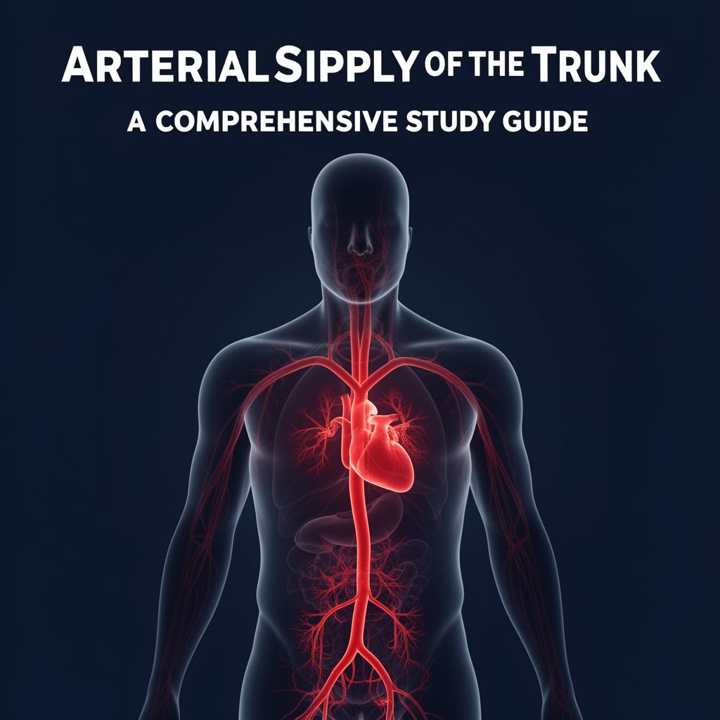
3.2. Abdominal Aorta 📊
The infra-diaphragmatic part of the descending aorta.
- Origin: Diaphragmatic hiatus.
- Termination: At L4 level, bifurcates into right and left common iliac arteries.
- Relations:
- Anterior (superior to inferior): Liver, peritoneum of bursa omentalis, stomach, pancreatic body, 3rd part of duodenum, left renal vein, mesenteric root, jejunal-ileal loops.
- Posterior: Vertebral column, left lumbar veins, left lumbar sympathetic nervous chain, thoracic duct, main lymphatic collector (Pequet).
- Right Lateral: Inferior vena cava.
- Left Lateral: Left kidney, left adrenal gland, left renal pelvis, left ureter.
- Branches:
- Terminal Branches: Right and left common iliac arteries, middle sacral artery (descends in the sub-aortic angle).
- Collateral Parietal Branches:
- Inferior diaphragmatic/phrenic arteries.
- Lumbar arteries (5 pairs, last pair from middle sacral artery).
- Collateral Visceral Branches:
- Coeliac trunk/artery of Haller.
- Superior mesenteric artery.
- Renal artery.
- Gonadal artery.
- Inferior mesenteric artery.
- Middle suprarenal artery.
3.3. Inferior Vena Cava (IVC) 📊
- Origin: Superior part of L5 vertebral body (union of right and left common iliac veins).
- Course: Ascends on the right side of the vertebral column.
- Termination: Crosses the diaphragmatic hiatus and opens into the inferior wall of the right atrium via an orifice guarded by the Eustachian valve.
- Relations:
- Anterior (superior to inferior): Liver, Winslow hiatus, 1st part of duodenum, pancreatic head, 3rd part of duodenum, right gonadal artery, mesenteric root, jejunal-ileal loops.
- Posterior: Right flank of vertebral column, lumbar arteries, right lumbar sympathetic chain.
- Right Lateral: Right adrenal gland, right kidney, right renal pelvis, right ureter, psoas muscle.
- Left Lateral: Abdominal aorta.
- Tributaries:
- Origin Tributaries: Right and left common iliac veins (left receives middle sacral vein).
- Parietal Tributaries: Inferior diaphragmatic veins, lumbar veins (5 pairs).
- Visceral Tributaries: Suprahepatic veins (2 large, 20 small), right and left renal veins, right middle suprarenal vein, right gonadal vein.
- 💡 Left gonadal vein typically drains into the left renal vein, then to IVC.
- Suprahepatic veins collect blood from the liver.
- Portal Vein: Formed by the union of the splenic vein, inferior mesenteric vein, and superior mesenteric vein.
3.4. Lymphatics of the Abdomen
- Collectors: Lymphatic vessels unite into two lumbar lymph collector tubes.
- Cisterna Chyli of Pequet: Receives lymph from lumbar collectors, continues superiorly as the thoracic duct.
- Lymph Nodes: Grouped into visceral and parietal.
- Parietal Lymph Nodes:
- Anterior lateral abdominal wall lymph nodes.
- Retroperitoneal parietal lymph nodes (lomboaortic): retrocaval, laterocaval, precaval, interaorticocaval groups.
- Visceral Lymph Nodes: Situated close to abdominal organs and along vessels (e.g., coeliac trunk and branches, superior and inferior mesenteric arteries and branches).
- Parietal Lymph Nodes:
3.5. Lumbar Sympathetic Chain
- Location: Two paravertebral sympathetic chain ganglia.
- Left side: Behind the aorta.
- Right side: Posterior to the inferior vena cava.
- Structure: Presents 5 pairs of sympathetic ganglia, communicating via intermediary columns.
- Continuity: Continuous superiorly with the thoracic sympathetic chain and inferiorly with the sacral sympathetic chain.
- Branches:
- Muscular.
- Vascular.
- Osseous.
- Communicating branches with lumbar spinal nerves.
- To the periaortic plexus (can be pre-aortic or inter-mesenteric).
- To the splanchnic nerves.
3.6. Solar Plexus (Coeliac Plexus) 💡
Also known as "the abdominal brain," it consists of ganglia and vegetative nervous fibers (afferent and efferent).
3.6.1. Ganglia
Typically three pairs of sympathetic ganglia, sometimes a fourth.
- Semilunar Ganglia (Right and Left): Each has anterior/posterior surfaces, concave superior/convex inferior borders, lateral/medial horns. Linked by a double bridge above and below the coeliac trunk.
- Aorticomesenteric Ganglia (Right and Left): Spherical, para-aortic, linked by a nervous bar below the inferior mesenteric artery origin.
- Aorticorenal Ganglia (Right and Left): Spherical, located beside renal artery emergencies.
- Retrorenal Ganglia of Hirschfeld (Right and Left): Small, inconstant, posterior to renal arteries, linked to aorticorenal ganglia.
3.6.2. Afferent Branches
Vegetative nervous fibers (sympathetic from thoracic sympathetic, parasympathetic from vagi nerves). Right phrenic nerve may also contribute.
- Splanchnic Nerves: Reach the superior border of the right semilunar ganglion near its lateral horn.
- Vagus Nerves:
- Initially descend on esophageal sides, then undergo torsion: left vagus anterior, right vagus posterior.
- Form a peri-esophageal vagal plexus.
- Give rise to anterior and posterior vagal trunks. The posterior vagal trunk (Delmas) contributes to the solar plexus.
- Solar Branches from Posterior Vagal Trunk:
- Right: Thick, reaches medial horn of right semilunar ganglion.
- Left: Thinner, reaches medial horn of left semilunar ganglion.
- Middle: Thinnest, descends on anterior aspect of aorta.
- Splanchnic Nerves (from Thoracic Sympathetic):
- Greater Splanchnic Nerve: Reaches the lateral horn of the semilunar ganglia.
- Lesser Splanchnic Nerve: Three branches: superior (to inferior border of semilunar ganglia), middle (to mesenteric ganglia), inferior (to aorticorenal ganglia).
- Imus Splanchnic Nerve: Inconstant, reaches aorticorenal ganglia.
3.6.3. Efferent Branches
Distributed to abdominal viscera, traveling as plexuses alongside abdominal vessels:
- Proper Coeliac Plexus: Gives off hepatic, lineal, and left gastric plexuses.
- Superior mesenteric plexus.
- Suprarenal plexus (paired).
- Renal plexus (paired).
- Gonadal plexus (paired).
- Inferior mesenteric plexus.
- Inferior diaphragmatic (phrenic) plexus (the only parietal plexus).