This study material has been compiled from various sources, including copy-pasted text from a document (likely a textbook or anatomical reference) and a lecture audio transcript.
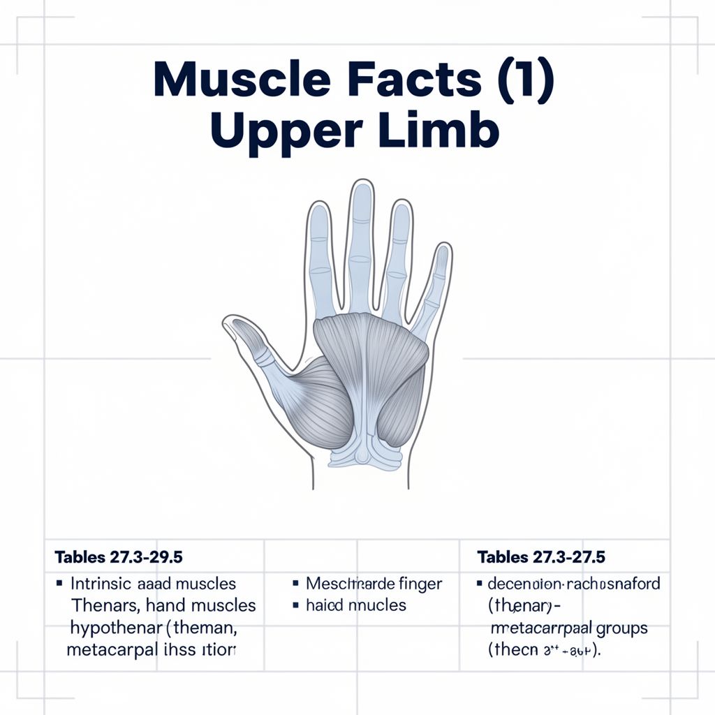
Intrinsic Muscles of the Hand: A Comprehensive Study Guide 🖐️
The human hand is a marvel of intricate design, allowing for both powerful grips and delicate, precise movements. This remarkable dexterity is largely attributed to the intrinsic muscles of the hand, which are located entirely within the hand itself. These muscles are crucial for fine motor control, manipulation of objects, and the overall functionality of the digits.
The intrinsic muscles are broadly categorized into three main groups based on their location and primary function:
- Thenar Muscles: Responsible for movements of the thumb.
- Hypothenar Muscles: Responsible for movements of the little finger.
- Metacarpal Muscles: Comprising the lumbricals and interossei, these muscles control the movements of the other digits (2nd through 5th fingers).
Let's delve into each group in detail.
1. Thenar Muscles: The Thumb's Movers 👍
The thenar eminence, the fleshy mound at the base of the thumb, houses a group of four muscles vital for thumb movement. These muscles enable the thumb's unique ability to oppose the other fingers, a key feature of human dexterity.
📚 Key Functions:
- Adduction: Moving the thumb towards the palm.
- Abduction: Moving the thumb away from the palm.
- Flexion: Bending the thumb at its joints.
- Opposition: Rotating the thumb to touch the tips of other fingers.
Individual Thenar Muscles:
1.1. Adductor Pollicis
- Origin:
- Transverse head: Palmar surface of the 3rd metacarpal.
- Oblique head: Capitate bone, bases of the 2nd and 3rd metacarpals.
- Insertion: Base of the proximal phalanx of the thumb, via the ulnar sesamoid bone.
- Innervation: Ulnar nerve (C8, T1).
- Action: ✅ Adduction of the thumb at the carpometacarpal (CMC) joint; flexion at the metacarpophalangeal (MCP) joint of the thumb.
1.2. Abductor Pollicis Brevis
- Origin: Scaphoid bone, trapezium, and flexor retinaculum.
- Insertion: Base of the proximal phalanx of the thumb, via the radial sesamoid bone.
- Innervation: Median nerve (C8, T1).
- Action: ✅ Abduction of the thumb at the CMC joint.
1.3. Flexor Pollicis Brevis
- Origin:
- Superficial head: Flexor retinaculum and trapezium.
- Deep head: Capitate bone and trapezium.
- Insertion: Base of the proximal phalanx of the thumb (superficial head via ulnar sesamoid, deep head via radial sesamoid).
- Innervation: Superficial head by the Median nerve (C8, T1); Deep head by the Ulnar nerve (C8, T1).
- Action: ✅ Flexion of the thumb at the CMC joint.
1.4. Opponens Pollicis
- Origin: Trapezium.
- Insertion: Radial border of the 1st metacarpal.
- Innervation: Median nerve (C8, T1).
- Action: ✅ Opposition of the thumb at the CMC joint, rotating it towards the palm to meet other fingers.
💡 These muscles work in concert to enable the thumb's critical functions in gripping and manipulation.
2. Hypothenar Muscles: The Little Finger's Control 🤏
The hypothenar eminence, located on the ulnar side of the palm at the base of the little finger, contains a group of muscles dedicated to the movements of the 5th digit. These muscles contribute to the hand's ability to cup the palm and perform fine adjustments with the little finger.
📚 Key Functions:
- Opposition: Drawing the little finger's metacarpal towards the palm.
- Flexion: Bending the little finger.
- Abduction: Moving the little finger away from the palm.
- Tightening: Providing protective tension to the palmar aponeurosis.
Individual Hypothenar Muscles:
2.1. Opponens Digiti Minimi
- Origin: Hook of hamate and flexor retinaculum.
- Insertion: Ulnar border of the 5th metacarpal.
- Innervation: Ulnar nerve (C8, T1).
- Action: ✅ Draws the 5th metacarpal in a palmar direction, contributing to opposition and cupping of the palm.
2.2. Flexor Digiti Minimi Brevis
- Origin: Hook of hamate and flexor retinaculum.
- Insertion: Base of the 5th proximal phalanx.
- Innervation: Ulnar nerve (C8, T1).
- Action: ✅ Flexion at the MCP joint of the little finger.
2.3. Abductor Digiti Minimi
- Origin: Pisiform bone.
- Insertion: Ulnar base of the 5th proximal phalanx and the dorsal digital expansion of the 5th digit.
- Innervation: Ulnar nerve (C8, T1).
- Action: ✅ Flexion and abduction at the MCP joint of the little finger; assists in extension of the PIP and DIP joints of the little finger.
2.4. Palmaris Brevis
- Origin: Ulnar border of the palmar aponeurosis.
- Insertion: Skin of the hypothenar eminence.
- Innervation: Ulnar nerve (C8, T1).
- Action: ✅ Tightens the palmar aponeurosis, providing a protective function to the palm's skin.
3. Metacarpal Muscles: Lumbricals and Interossei ✍️
These muscles are located between the metacarpal bones and are crucial for the precise movements of the 2nd through 5th digits, enabling fine motor skills like writing, playing instruments, and manipulating small objects.
3.1. Lumbricals
There are four lumbrical muscles, unique in that they originate from tendons and insert into tendons/expansions.
- Origin: Tendons of the flexor digitorum profundus.
- 1st & 2nd Lumbricals: Radial sides of the flexor digitorum profundus tendons.
- 3rd & 4th Lumbricals: Bipennate, from the medial and lateral sides of the flexor digitorum profundus tendons.
- Insertion: Dorsal digital expansion (dde) of the 2nd to 5th digits.
- Innervation:
- 1st & 2nd Lumbricals: Median nerve (C8, T1).
- 3rd & 4th Lumbricals: Ulnar nerve (C8, T1).
- Action: ✅ Flexion at the MCP joints and extension at the proximal and distal interphalangeal (IP) joints of the 2nd to 5th digits. This action is often described as the "number-making" or "waiter's" position, where the MCP joints are flexed and the IP joints are extended.
3.2. Dorsal Interossei
There are four dorsal interossei muscles, each bipennate and responsible for abducting the fingers.
- Origin: Adjacent sides of the metacarpals.
- 1st: 1st and 2nd metacarpals.
- 2nd: 2nd and 3rd metacarpals.
- 3rd: 3rd and 4th metacarpals.
- 4th: 4th and 5th metacarpals.
- Insertion: Dorsal digital expansion (dde) and the radial/ulnar side of the proximal phalanx of the 2nd to 4th digits.
- 1st: Radial side of 2nd proximal phalanx.
- 2nd: Radial side of 3rd proximal phalanx.
- 3rd: Ulnar side of 3rd proximal phalanx.
- 4th: Ulnar side of 4th proximal phalanx.
- Innervation: Ulnar nerve (C8, T1).
- Action: ✅ Flexion at the MCP joints, extension at the proximal and distal IP joints, and abduction of the 2nd to 4th digits away from the 3rd digit.
3.3. Palmar Interossei
There are three palmar interossei muscles, responsible for adducting the fingers.
- Origin:
- 1st: Ulnar side of the 2nd metacarpal.
- 2nd: Radial side of the 4th metacarpal.
- 3rd: Radial side of the 5th metacarpal.
- Insertion: Dorsal digital expansion (dde) and the base of the proximal phalanx of the 2nd, 4th, and 5th digits.
- Innervation: Ulnar nerve (C8, T1).
- Action: ✅ Flexion at the MCP joints, extension at the proximal and distal IP joints, and adduction of the 2nd, 4th, and 5th digits towards the 3rd digit.
⚠️ Important Note on Innervation:
While the median nerve primarily innervates the thenar muscles (except for the deep head of flexor pollicis brevis and adductor pollicis), and the ulnar nerve primarily innervates the hypothenar and interossei muscles, there is a shared innervation for the lumbricals (1st and 2nd by median, 3rd and 4th by ulnar) and a dual innervation for the flexor pollicis brevis. This complex innervation pattern highlights the intricate control mechanisms of the hand.
Understanding these intrinsic muscles, their origins, insertions, innervations, and actions is fundamental to comprehending the complex motor control and functional capabilities of the human hand.