Heme and Cholesterol Metabolism: Essential Biochemical Pathways - kapak
Bilim#heme#cholesterol#metabolism#biochemistry

Heme and Cholesterol Metabolism: Essential Biochemical Pathways

Explore the vital biochemical pathways of heme and cholesterol, from their intricate synthesis and diverse functions to their regulation, catabolism, and clinical implications in human health.

ilaydadyaliiJanuary 22, 2026 ~13 dk toplam
01

Flash Kartlar

25 kart

Karta tıklayarak çevir. ← → ile gez, ⎵ ile çevir.

1 / 25
Tüm kartları metin olarak gör
  1. 1. Describe the fundamental structural characteristics of heme.

    Heme is characterized by a heterocyclic ring structure composed of four pyrrole rings interconnected by carbon (methenyl) bridges. At the core of this porphyrin structure, a metal atom is chelated to the nitrogen atoms of the pyrrole units. In heme, this central metal atom is specifically iron.

  2. 2. What is the key difference in the central metal atom between heme and chlorophyll?

    The key difference lies in the central metal atom chelated within their porphyrin structures. In heme, the central atom is iron, which is crucial for its oxygen-binding capabilities. Conversely, in chlorophyll, the central atom is magnesium, essential for its role in photosynthesis.

  3. 3. Explain two primary functions of heme related to oxygen.

    Heme is indispensable for oxygen transport in hemoglobin, allowing red blood cells to carry oxygen from the lungs to tissues. It is also vital for oxygen storage in myoglobin, particularly in muscle tissues, providing an oxygen reserve for metabolic activity. These roles highlight heme's critical involvement in cellular respiration and energy production.

  4. 4. List three other vital functions of heme beyond oxygen transport and storage.

    Beyond oxygen roles, heme serves as a prosthetic group for cytochrome P450 enzymes, which are involved in detoxification and metabolism. It acts as a critical reservoir of iron within the body, and functions as an electron shuttle for enzymes within the electron transport chain, fundamental to cellular respiration. Heme also plays roles in signal transduction, regulating circadian rhythms, and cellular differentiation.

  5. 5. Which are the primary organs responsible for heme synthesis?

    The primary organs responsible for heme synthesis are the liver and the bone marrow. The liver synthesizes heme for various hemoproteins, while the bone marrow is crucial for producing heme for hemoglobin in developing red blood cells. These two sites account for the majority of heme production in the body.

  6. 6. In which type of red blood cells is heme synthesized, and why is this distinction important?

    Heme is not synthesized in mature red blood cells, which lack mitochondria. Instead, it is synthesized in erythroid cells, particularly immature erythrocytes known as reticulocytes. This distinction is important because mature red blood cells are terminally differentiated and primarily function in oxygen transport, while reticulocytes still possess the necessary machinery for heme production before fully maturing.

  7. 7. How does heme influence globin chain synthesis in erythroid cells?

    In erythroid cells, heme actively stimulates the protein synthesis of globin chains. This coordinated synthesis is essential for the proper formation of hemoglobin, as heme and globin chains must combine in specific ratios. The accumulation of heme in these cells favors globin chain synthesis, which is crucial for erythroblast maturation and efficient oxygen transport.

  8. 8. Describe the initial, rate-limiting step of heme synthesis, including its location and key reactants.

    The initial and rate-limiting step of heme synthesis occurs within the mitochondria. In this reaction, d-aminolevulinic acid synthase (ALA synthase) catalyzes the condensation of succinyl-CoA and glycine. This process forms S-Aminolevulinate (ALA), along with CoA and carbon dioxide, marking the committed step in the heme biosynthesis pathway.

  9. 9. What enzyme catalyzes the initial step of heme synthesis, and what is its significance?

    The enzyme d-aminolevulinic acid synthase, or ALA synthase, catalyzes the initial step of heme synthesis. This enzyme is highly significant because it controls the rate-limiting step of the entire pathway. Its activity is tightly regulated, making it a primary control point for overall heme production in the body.

  10. 10. How is heme synthesis primarily regulated in erythroid cells and the liver?

    Heme synthesis is tightly regulated, primarily in erythroid cells and the liver, by controlling the activity of ALA synthase. Heme itself acts as a potent feedback inhibitor, ensuring that production matches demand. This regulation prevents the overproduction or underproduction of heme, which can have significant physiological consequences.

  11. 11. Explain how heme acts as a feedback inhibitor of ALA synthase.

    Heme acts as a potent feedback inhibitor of ALA synthase through several mechanisms. As heme levels rise, it directly suppresses the enzyme's activity. This inhibition occurs by blocking the transport of ALA synthase from the cytoplasm to the mitochondria and by inhibiting the de novo synthesis of the enzyme itself, thereby reducing its overall availability.

  12. 12. What happens to heme after it has fulfilled its biological roles, and what is preserved?

    Once heme has fulfilled its biological roles, it cannot be re-used or recycled in its original form. Therefore, it undergoes a catabolic process where it is transformed into bilirubin and subsequently excreted from the body. Crucially, the iron atom from heme is preserved and recycled, allowing it to be reused for new heme synthesis or other iron-dependent processes.

  13. 13. What is the primary source of heme that undergoes catabolism to form bilirubin?

    The primary source of heme that undergoes catabolism to form bilirubin is the hemoglobin from aged red blood cells. This accounts for approximately 85-90% of the total heme degraded. The remaining 10-15% comes from the catabolism of other hemoproteins like myoglobin, cytochromes, and heminic enzymes.

  14. 14. What is the typical lifespan of an erythrocyte, and how are aged ones removed from the body?

    The typical lifespan of an erythrocyte (red blood cell) is approximately 120 days. Aged red blood cells are recognized and phagocytized by macrophages within the endothelial reticulum system, primarily in the spleen, liver, and bone marrow. This process is known as hemocateresis, ensuring the removal of old and damaged cells from circulation.

  15. 15. Which enzyme initiates heme degradation, and what does it catalyze?

    Heme degradation is initiated by heme oxygenase, a microsomal enzyme. This enzyme catalyzes the region-specific opening of the heme ring. This crucial step leads to the formation of biliverdin, carbon monoxide, and the release of the iron atom, marking the beginning of heme's conversion into bile pigments.

  16. 16. What are the main products formed during the heme oxygenase reaction?

    The heme oxygenase reaction, which initiates heme degradation, produces three main products. These are biliverdin, a green pigment; carbon monoxide (CO); and a free iron atom. This process requires molecular oxygen and reducing equivalents provided by NADPH-cytochrome P450 reductase.

  17. 17. Describe the process of bilirubin conjugation in the liver.

    After bilirubin is released into the bloodstream and transported to the liver bound to serum albumin, it undergoes conjugation. Within hepatocyte microsomes, bilirubin is conjugated with UDP-glucuronic acid. This reaction forms a water-soluble product called bilirubindiglucuronide, which is essential for its excretion.

  18. 18. Explain the fate of conjugated bilirubin in the intestine, leading to the formation of urobilinogen and stercobilin.

    Conjugated bilirubin (bilirubindiglucuronide) is actively secreted into the bile and enters the intestine. There, bacterial flora retransforms it into bilirubin and then into degradation products, including urobilinogen. A portion of urobilinogen is reabsorbed and excreted by the kidneys as urobilin, while the majority is converted to stercobilin, which gives feces its brown color.

  19. 19. Define hyperbilirubinemia and describe its clinical manifestation.

    Hyperbilirubinemia is defined as a condition where bilirubin levels in the blood exceed 2 mg/dL. Its primary clinical manifestation is jaundice, which is characterized by a yellow coloring of the sclerae (whites of the eyes) and the skin. This yellow discoloration occurs due to the accumulation of bilirubin in tissues.

  20. 20. What causes pre-hepatic (hemolytic) jaundice?

    Pre-hepatic, or hemolytic, jaundice is caused by an excessive breakdown of red blood cells, leading to an overproduction of bilirubin. This overwhelms the liver's capacity to conjugate and excrete bilirubin. Consequently, there is an accumulation of indirect, or unconjugated, bilirubin in the blood, resulting in the characteristic yellow discoloration.

  21. 21. Describe the fundamental structural characteristics of cholesterol.

    Cholesterol is a lipid, specifically a sterol, characterized by its fundamental perhydro-cyclo-pentano-phenanthrene structure, which consists of four fused rings. It features a hydroxyl group at carbon 3, a double bond between carbon 5 and 6, an eight-carbon side chain at carbon 17, and methyl groups at carbons 10 and 13. This unique structure gives it a polar head and a non-polar tail.

  22. 22. How does cholesterol's structure enable its role in cell membranes?

    Cholesterol's unique structure, with a polar hydroxyl group head and a non-polar, flexible tail, allows it to interact effectively with cell membranes. Its polar head interacts with the aqueous environment and the polar heads of phospholipids, while its rigid ring system and non-polar tail embed within the hydrophobic core of the membrane. This integration helps maintain membrane fluidity and stability.

  23. 23. List two types of essential molecules for which cholesterol serves as a precursor.

    Cholesterol serves as a crucial precursor for several essential molecules. It is the building block for all steroid hormones, including sex hormones like testosterone, estrogens, and progesterone, as well as stress hormones such as cortisol. Additionally, cholesterol is a precursor for bile salts, which are necessary for the digestion and emulsification of dietary fats.

  24. 24. Differentiate between the primary roles of cholesterol and triglycerides in the body.

    Triglycerides primarily serve as the body's mechanism for storing unused calories for future energy needs, acting as a long-term energy reserve. In contrast, cholesterol is a lipid used to build and repair cell membranes, maintaining their fluidity and integrity. It also functions as a precursor for vital hormones and bile acids, highlighting its structural and signaling roles rather than energy storage.

  25. 25. Explain the difference between HDL and LDL cholesterol, including their common names and functions.

    HDL (High-Density Lipoprotein) is often called 'good' cholesterol because it contains more protein than cholesterol. Its function is to transport cholesterol from peripheral tissues back to the liver for elimination, thus protecting against plaque buildup. LDL (Low-Density Lipoprotein) is known as 'bad' cholesterol, containing more cholesterol than protein. It deposits cholesterol in damaged blood vessels, contributing to plaque formation and increasing the risk of coronary artery disease.

02

Detaylı Özet

8 dk okuma

Tüm konuyu derinlemesine, başlık başlık.

Comprehensive Study Material: Heme, Cholesterol, and Carbohydrate Metabolism

Source Information: This study material has been compiled from a lecture audio transcript and various copy-pasted text sources (personal notes, PDF/PowerPoint texts).


Introduction

This study guide explores the fundamental biochemical pathways and clinical significance of heme, cholesterol, and carbohydrates. These molecules are vital for energy production, structural integrity, signaling, and overall cellular function. Understanding their synthesis, degradation, and regulation is crucial for comprehending human physiology and disease.


I. Heme Metabolism

Heme is a critical prosthetic group found in various proteins, essential for oxygen transport, electron transfer, and drug metabolism.

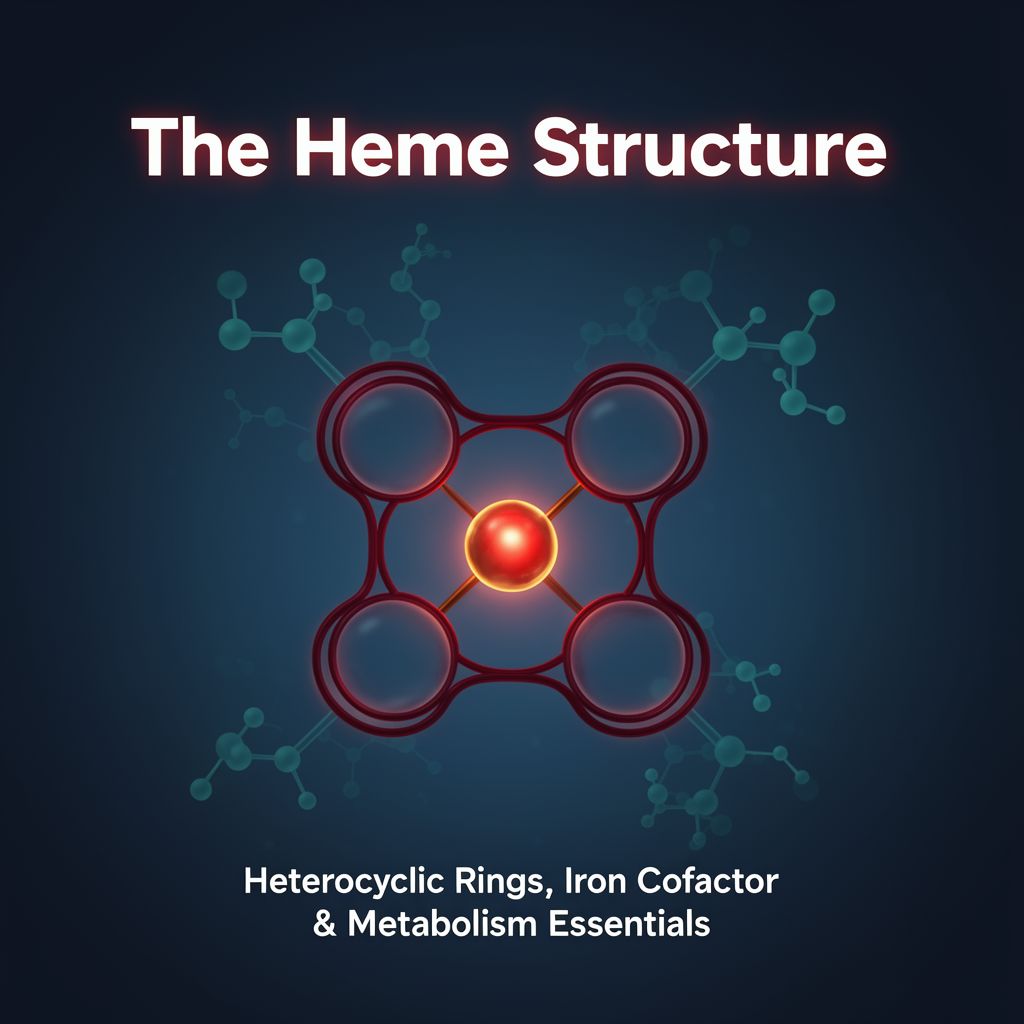
A. Heme Structure & Functions 📚

Heme features a heterocyclic ring structure composed of four pyrrole rings joined by methenyl bridges. A metal atom is chelated at its center to the nitrogen atoms of the pyrrole units.

  • In Heme: The central metal atom is iron (Fe).
  • In Chlorophyll: The central metal atom is magnesium (Mg).

Key Functions of Heme: ✅ Oxygen transport (hemoglobin) and storage (myoglobin) ✅ Prosthetic group for cytochrome P450 enzymes ✅ Iron reservoir ✅ Electron shuttle in the electron transport chain (cellular respiration) ✅ Signal transduction (e.g., circadian rhythms, antioxidant response) ✅ Cellular differentiation and proliferation ✅ Nitric oxide synthase activity

B. Heme Synthesis Pathway 🧬

Heme synthesis primarily occurs in the liver and bone marrow, specifically in erythroid cells (not mature red blood cells).

  • Initial Step (Mitochondria): 1️⃣ Succinyl-CoA + Glycine → S-Aminolevulinate (ALA)
    • Catalyzed by d-aminolevulinic acid synthase (ALA synthase).
    • Involves condensation and decarboxylation.
    • ALA is then exported to the cytoplasm.
  • Cytoplasmic Steps:
    • 2️⃣ 2 ALA → Porphobilinogen (PBG) + 2 H₂O
      • Catalyzed by ALA dehydratase (also Porphobilinogen Synthase).
      • Highly sensitive to heavy metal inhibition (e.g., lead poisoning).
    • 3️⃣ 4 PBG → Linear Hydroxymethylbilane (HMB)
      • Catalyzed by Porphobilinogen Deaminase (Hydroxymethylbilane Synthase), releasing 4 ammonia molecules.
    • 4️⃣ HMB → Uroporphyrinogen III → Coproporphyrinogen III
      • Uroporphyrinogen Decarboxylase converts Uroporphyrinogen III to Coproporphyrinogen III, which enters the mitochondria.
  • Mitochondrial Steps (Final):
    • 5️⃣ Coproporphyrinogen III → Protoporphyrinogen IX → Protoporphyrin IX
      • Involves oxidation steps catalyzed by enzymes like Protoporphyrinogen IX Oxidase.
    • 6️⃣ Protoporphyrin IX + Fe²⁺ → Heme
      • Catalyzed by Ferrochelatase.

C. Regulation of Heme Synthesis ⚖️

The rate-limiting step is controlled by ALA synthase.

  • Feedback Inhibition: Heme directly inhibits ALA synthase activity.
  • Transport Inhibition: Heme blocks ALA synthase transport from cytoplasm to mitochondria.
  • Gene Expression: Heme suppresses the synthesis of ALA synthase.
  • Erythroid Cells: Heme stimulates globin chain synthesis, and erythropoietin stimulates heme production, crucial for erythroblast maturation.

D. Heme Catabolism & Bilirubin ♻️

Heme cannot be reused; it is degraded, but its iron is preserved.

  • Process: Aged red blood cells (~120 days) are phagocytized by macrophages in the spleen, liver, and bone marrow. Hemoglobin is split into globin and heme.
  • Heme Degradation:
    • 1️⃣ Heme → Biliverdin + CO + Fe²⁺
      • Catalyzed by Heme Oxygenase (microsomal enzyme), requiring O₂ and NADPH.
    • 2️⃣ Biliverdin → Bilirubin
      • Catalyzed by Biliverdin Reductase.
  • Bilirubin Transport & Excretion:
    • Bilirubin (unconjugated/indirect) binds to serum albumin and travels to the liver.
    • In hepatocytes, it's conjugated with UDP-glucuronic acid by UDP-glucuronyltransferase to form water-soluble bilirubindiglucuronide (conjugated/direct).
    • Conjugated bilirubin is secreted into bile.
    • In the intestine, bacterial flora convert it to urobilinogen.
    • Urobilinogen is mostly converted to stercobilin (colors feces brown) or reabsorbed and excreted as urobilin in urine (yellow pigment).

E. Clinical Aspects of Heme Metabolism 🏥

  • Porphyrias: Genetic disorders due to enzyme deficiencies in heme synthesis, leading to accumulation of heme precursors.
    • Acute Intermittent Porphyria: Mutation in Hydroxymethylbilane Synthase, accumulating ALA and PBG.
    • Porphyria Cutanea Tarda: Deficiency in Uroporphyrinogen Decarboxylase.
  • Lead Poisoning: Lead inhibits several heme synthesis enzymes, notably ALA dehydratase and Ferrochelatase.
  • Jaundice (Hyperbilirubinemia): Yellow discoloration of skin/sclerae (bilirubin > 2 mg/dL).
    • Pre-hepatic (Hemolytic): Excess indirect bilirubin (e.g., acute hemolytic anemia).
    • Hepatic (Hepatocellular): Both direct and indirect bilirubin (e.g., liver damage).
    • Post-hepatic (Cholestatic): Excess direct bilirubin (e.g., bile duct obstruction).

II. Cholesterol Metabolism

Cholesterol is a vital lipid, serving as a structural component of cell membranes and a precursor for essential biomolecules.

A. Cholesterol Structure & Functions 🔬

  • Structure: Perhydro-cyclo-pentano-phenanthrene (four fused rings) with a hydroxyl group at C3, a double bond at C5-C6, an 8-carbon side chain at C17, and methyl groups at C10 and C13. It has a polar head, rigid ring system, and non-polar tail.
  • Functions: ✅ Cell membrane fluidity and integrity (especially nervous cells). ✅ Precursor for bile salts (fat digestion). ✅ Precursor for steroid hormones (sex hormones, stress hormones). ✅ Precursor for fat-soluble vitamins (D, K, A, E).

B. Cholesterol vs. Triglycerides & Lipoproteins 📊

  • Triglycerides: Store unused calories for energy. High levels linked to obesity, metabolic syndrome.
  • Cholesterol: Builds cell membranes, produces hormones.
  • Lipoproteins: Transport cholesterol in blood.
    • HDL ("Good" Cholesterol): High-Density Lipoprotein. More protein than cholesterol. Transports cholesterol from tissues to liver for excretion (reverse cholesterol transport). Higher HDL = lower coronary artery disease (CAD) risk.
    • LDL ("Bad" Cholesterol): Low-Density Lipoprotein. More cholesterol than protein. Deposits cholesterol in blood vessels, leading to plaque buildup. Higher LDL = increased CAD risk.

C. Cholesterol Biosynthesis Pathway 🧪

  • Location: Primarily liver and intestine; occurs in cytosol and mitochondria.
  • Precursor: Acetyl-CoA. Requires ATP and NADPH.
  • Key Steps:
    1. Acetyl-CoA → HMG-CoA: Two Acetyl-CoA condense to Acetoacetyl-CoA, then react with a third Acetyl-CoA to form HMG-CoA.
    2. HMG-CoA → Mevalonate: Rate-limiting step, catalyzed by HMG-CoA Reductase (HMGR), requiring 2 NADPH.
    3. Mevalonate → Isopentenyl Pyrophosphate (IPP) & Dimethylallyl Pyrophosphate (DMAPP): Involves phosphorylation and decarboxylation. These are 5-carbon isoprene units.
    4. IPP + DMAPP → Squalene: IPP and DMAPP condense to Geranyl Pyrophosphate (GPP, 10C), then to Farnesyl Pyrophosphate (FPP, 15C). Two FPP molecules join to form Squalene (30C).
    5. Squalene → Lanosterol → Cholesterol: Squalene undergoes cyclization (via Squalene Epoxidase and Lanosterol Synthase) to Lanosterol, which is then converted to cholesterol through 19 additional reactions.

D. Regulation of Cholesterol Synthesis ⚙️

HMGR is the primary regulatory enzyme.

  • Feedback Inhibition: High cholesterol inhibits HMGR activity and promotes its degradation.
  • Transcriptional Regulation:
    • SREBP (Sterol Regulatory Element-Binding Protein): A transcription factor that activates HMGR gene expression.
    • SCAP (SREBP Cleavage-Activating Protein) & INSIG proteins: When sterol levels are high, SCAP-SREBP complex remains in the ER, bound to INSIG, preventing SREBP activation.
    • Low Sterols: SCAP-SREBP complex moves to Golgi, where SREBP is proteolytically cleaved by Site-1 and Site-2 Proteases (S1P, S2P). The active SREBP fragment enters the nucleus, activating HMGR gene transcription.
  • Short-Term Regulation:
    • AMPK (AMP-activated protein kinase): Phosphorylates and inhibits HMGR when cellular energy (ATP) is low, reducing energy-demanding cholesterol synthesis.
  • Pharmaceutical Regulation:
    • Statins: Competitively inhibit HMGR, mimicking mevalonate, thereby reducing cholesterol synthesis and lowering blood cholesterol.

E. Cholesterol Transport & Esterification 🚚

  • Esterification: Cholesterol is converted to cholesterol esters (more hydrophobic) for storage and transport.
    • ACAT (Acyl-CoA-Cholesterol Acyltransferase): Catalyzes esterification in cells, using Acyl-CoA. ACAT1 (all tissues), ACAT2 (intestine).
    • LCAT (Lecithin-Cholesterol Acyltransferase): Acts on HDL, using lecithin to esterify cholesterol, trapping it within HDL.
  • Reverse Cholesterol Transport: HDL picks up cholesterol from peripheral tissues, LCAT esterifies it, and HDL delivers cholesterol esters to the liver via SR-B1 receptor and ABC transporters.

III. Carbohydrate Biochemistry

Carbohydrates are polyfunctional compounds crucial for energy, structure, and cellular communication.

A. Carbohydrate Basics & Functions 💡

  • Definition: Polyhydroxyaldehydes or polyhydroxyketones. "Sugars" are water-soluble and sweet carbohydrates.
  • Functions: ✅ Primary energy source (glucose). ✅ Energy storage (starch in plants, glycogen in animals). ✅ Structural components (cellulose in plants, chitin in insects, bacterial cell walls). ✅ Components of DNA and RNA. ✅ Cell communication and recognition (glycobiology, glycomics).

B. Monosaccharide Classification & Stereochemistry 🔄

  • Monosaccharides: Simplest carbohydrates, primary energy source, building blocks.
    • Classification by Carbon Atoms: Triose (3C), Tetrose (4C), Pentose (5C), Hexose (6C, e.g., glucose).
    • Classification by Carbonyl Group: Aldoses (aldehyde at C1), Ketoses (ketone at C2).
  • Stereochemistry:
    • Chiral Carbon: Carbon bonded to four different groups, creating enantiomers (non-superimposable mirror images).
    • D/L Configuration: Determined by the -OH group on the chiral carbon farthest from the functional group. D-form (OH on right) is most common in nature.
    • Epimers: Sugars differing in configuration at only one chiral carbon (e.g., glucose and mannose are C2 epimers).

C. Ring Formation & Anomers 💍

  • Cyclization: 5- and 6-carbon monosaccharides form rings in solution via intramolecular hemiacetal (aldoses) or hemiketal (ketoses) formation.
    • Pyranose: Six-membered ring (e.g., glucose forms glucopyranose).
    • Furanose: Five-membered ring (e.g., fructose forms fructofuranose).
  • Anomers: New chiral center formed at the anomeric carbon (C1 for aldoses, C2 for ketoses) upon cyclization.
    • Alpha (α) Anomer: -OH on anomeric carbon is opposite to the -CH₂OH group (below the plane in Haworth projection).
    • Beta (β) Anomer: -OH on anomeric carbon is on the same side as the -CH₂OH group (above the plane).
  • Mutarotation: Equilibrium between α and β anomers in solution, interconverting via the open-chain form, leading to a stable optical rotation.

D. Monosaccharide Derivatives

Simple sugars can be modified by adding or changing functional groups.

  • Phosphorylation: Addition of phosphate groups (e.g., glucose 6-phosphate). Traps sugars inside cells.
  • Oxidation: Conversion of -OH or aldehyde groups to acids.
  • Amination: Replacement of -OH with amino groups (e.g., glucosamine).
  • Dehydrogenation: Removal of hydrogen atoms. These derivatives are crucial for cell recognition, signaling, and metabolism.

IV. Related Metabolic & Clinical Concepts

A. Fatty Acid Metabolism Regulation ⚖️

  • Acetyl-CoA Carboxylase (ACC): Key enzyme controlling fatty acid synthesis.
    • Hormonal Regulation: Insulin activates ACC (synthesis), Glucagon/Epinephrine inhibit ACC (breakdown).
    • Allosteric Regulation: Citrate activates ACC (abundant energy), Palmitoyl-CoA inhibits ACC (sufficient fatty acids).
    • Energy Status: AMPK phosphorylates and inactivates ACC when energy is low (high AMP).
  • Fed State: Insulin promotes storage, inhibits fat breakdown, stimulates malonyl-CoA (inhibits fatty acid entry into mitochondria for oxidation).
  • Starvation: Glucagon/Epinephrine activate lipase, inactivate ACC, reduce malonyl-CoA, increasing fatty acid transport into mitochondria for beta-oxidation.

B. Neurodegenerative Diseases & Metabolism (MS, ALS) 🧠

Mitochondrial dysfunction and oxidative stress are common features in neurodegenerative diseases.

  • Mitochondrial Dysfunction: Problems in Krebs cycle, electron transport chain, oxidative phosphorylation lead to ATP imbalance.
  • Reactive Oxygen Species (ROS) & Reactive Nitrogen Species (RNS): Highly reactive molecules causing oxidative/nitrosative stress, damaging lipids, proteins, DNA. Imbalance contributes to disease progression.
  • Multiple Sclerosis (MS): Demyelinating disease. ROS/RNS contribute to myelin and oligodendrocyte damage. Mitochondrial dysfunction plays a role in progression.
  • Amyotrophic Lateral Sclerosis (ALS): Motor neuron disease. Characterized by oxidative/nitrosative stress and mitochondrial dysfunction. Metabolic alterations include altered NAA levels, amino acid changes, reduced antioxidants (alpha/gamma-tocopherol, GSH), and elevated oxidative damage markers (MDA, nitrite/nitrate).

Kendi çalışma materyalini oluştur

PDF, YouTube videosu veya herhangi bir konuyu dakikalar içinde podcast, özet, flash kart ve quiz'e dönüştür. 1.000.000+ kullanıcı tercih ediyor.

Sıradaki Konular

Tümünü keşfet
Understanding the TCA Cycle: Enzymes and Regulation

Understanding the TCA Cycle: Enzymes and Regulation

Explore the intricacies of the Tricarboxylic Acid (TCA) cycle, its key enzymes, regulatory mechanisms, and clinical implications, providing a detailed overview of this central metabolic pathway.

Özet 25 15
Biochemistry and Cellular Structure: Foundations of Life

Biochemistry and Cellular Structure: Foundations of Life

This summary explores biochemistry as the chemical basis of life, detailing metabolic processes, cellular organization, and the specialized functions of prokaryotic and eukaryotic cell components.

7 dk Özet 25 15
Unlocking Life's Chemistry: An Intro to Biochemistry

Unlocking Life's Chemistry: An Intro to Biochemistry

Explore the fascinating world of biochemistry, understanding the chemical processes and molecules that make life possible, from DNA to energy production.

Özet 25 15
Cellular Organization and Molecular Foundations

Cellular Organization and Molecular Foundations

Explore the intricate world of eukaryotic cells, from their diverse numbers, shapes, and sizes to the fundamental chemical elements and organic molecules that form the basis of life.

10 dk Özet 25 15
Riboflavin and Niacin: Essential B Vitamins

Riboflavin and Niacin: Essential B Vitamins

Explore the absorption, functions, metabolism, and clinical aspects of Riboflavin (Vitamin B2) and Niacin (Vitamin B3), including their roles as coenzymes and implications of deficiency and toxicity.

Özet 15
Cellular Respiration and Energy Metabolism

Cellular Respiration and Energy Metabolism

An academic summary detailing cellular respiration, its stages, types, and the broader context of energy metabolism, including nutrient processing and energy balance.

6 dk Özet 25 15
Common Characteristics of Living Organisms

Common Characteristics of Living Organisms

An academic summary detailing the fundamental characteristics shared by all living organisms, including cellular organization, metabolism, homeostasis, reproduction, growth, response to stimuli, and adaptation.

5 dk Özet 25 15
Common Characteristics of Living Organisms

Common Characteristics of Living Organisms

An academic summary detailing the fundamental properties that define living organisms, including cellular organization, metabolism, reproduction, growth, and adaptation, for foundational biological understanding.

6 dk Özet 25 15