Sesli Özet
18 dakikaKonuyu otobüste, koşarken, yolda dinleyerek öğren.
Sesli Özet
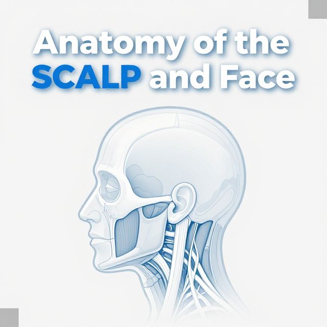
Anatomy of the SCALP and Face
Flash Kartlar
25 kartKarta tıklayarak çevir. ← → ile gez, ⎵ ile çevir.
Tüm kartları metin olarak gör
1. What is the anatomical definition of the SCALP?
The SCALP is defined as the skin and the underlying subcutaneous tissue covering the calvaria. It extends between the superior nuchal lines posteriorly and the supraorbital margins anteriorly. This region is crucial for protecting the skull and brain.
2. List the five layers of the SCALP from superficial to deep.
The five layers of the SCALP are: Skin, Connective tissue, Aponeurosis, Loose areolar tissue, and Pericranium. Each layer has distinct characteristics and functions, contributing to the overall structure and resilience of the scalp.
3. Describe the key characteristics of the Skin layer of the SCALP.
The Skin layer of the SCALP is notably hairy and thick. It contains numerous sweat and sebaceous glands, which contribute to its protective and thermoregulatory functions. Furthermore, it is richly supplied with arteries, veins, and lymphatic vessels, ensuring its metabolic needs are met.
4. What are the main features of the Connective tissue layer of the SCALP?
The Connective tissue layer is formed by thick and dense connective tissue. This layer is significant because it houses the sensory nerves of the scalp, providing sensation. Additionally, it boasts a rich vascular supply, which is essential for nourishing the scalp tissues.
5. Explain the structure and function of the Aponeurosis layer of the SCALP.
The Aponeurosis is a dense tendinous structure within the SCALP. Its primary function is to serve as the attachment point for the muscles that form the epicranius muscle. This attachment is crucial for movements of the scalp and forehead, contributing to facial expressions.
6. What is unique about the Loose areolar tissue layer of the SCALP, especially regarding its vascular components?
The Loose areolar tissue layer has a characteristic spongy structure. It contains a relatively small number of arteries but crucially houses emissary veins. These emissary veins are vital connections that extend between the superficial veins of the scalp and the diploic veins within the skull bones, serving as important drainage pathways.
7. Describe the Pericranium layer of the SCALP.
The Pericranium is the deepest layer of the SCALP. It is essentially the periosteum of the skull bones, meaning it's a dense connective tissue membrane that covers the outer surface of the skull. This layer provides nourishment and protection to the underlying bone.
8. Which layers of the SCALP are collectively referred to as the 'scalp proper' and why?
The first three layers of the SCALP—the Skin, Connective tissue, and Aponeurosis—are collectively referred to as the 'scalp proper.' This is because these layers are firmly attached together. During head injuries or surgical openings of the cranium, they often separate as a single, cohesive unit.
9. Name the main arteries supplying the SCALP that originate from the external carotid artery.
The main arteries supplying the SCALP that originate from the external carotid artery are the Occipital artery, the Posterior auricular artery, and the Superficial temporal artery. These vessels provide a significant portion of the blood supply to the scalp, ensuring its metabolic needs are met.
10. Which arteries supply the SCALP and are branches of the ophthalmic artery?
The arteries supplying the SCALP that are branches of the ophthalmic artery (which itself is a branch of the internal carotid artery) are the Supratrochlear artery and the Supraorbital artery. These arteries contribute to the anterior blood supply of the scalp, particularly in the forehead region.
11. List the regional lymph nodes into which the lymphatics of the SCALP drain.
The lymphatics of the SCALP drain into several regional lymph nodes, including the Submental, Submandibular, Parotid, Mastoid, Retroauricular, and Occipital lymph nodes. These nodes then primarily drain into the deep cervical lymph nodes, forming part of the larger lymphatic system.
12. Which cranial nerve branches provide sensory innervation to the SCALP anterior to the auricle?
Sensory innervation to the SCALP anterior to the auricle is primarily received by three branches of the fifth cranial nerve (CN V), also known as the trigeminal nerve. These branches are the Ophthalmic nerve, Maxillary nerve, and Mandibular nerve, covering distinct regions of the anterior scalp.
13. Which nerves provide sensory innervation to the SCALP posterior to the auricle?
Posterior to the auricle, sensory innervation to the SCALP is supplied by the dorsal rami of the cervical spinal nerves, specifically C2 and C3. These include the Greater occipital nerve and the Lesser occipital nerve, which cover the posterior and inferoposterior parts of the scalp.
14. Name the main veins involved in the drainage of the SCALP.
The veins of the SCALP generally follow the arterial supply and drain into the Occipital vein, Posterior auricular vein, Superficial temporal vein, Supratrochlear vein, and Supraorbital vein. These veins then contribute to larger venous systems, ultimately returning blood to the heart.
15. Which cranial nerve innervates all the muscles of facial expression?
All the muscles of facial expression are innervated by the seventh cranial nerve, known as the facial nerve (CN VII). Specifically, the nerve fibers responsible for this innervation are classified as Special Visceral Efferent (SVE) fibers. This unique innervation allows for the wide range of human facial expressions.
16. What is a key anatomical characteristic regarding fascia in the face?
A key anatomical characteristic of the face is the absence of deep fascia, which is a dense fibrous connective tissue layer found in many other parts of the body. Instead, the superficial fascia in the face is present and is notably thicker in certain regions, allowing for the mobility of facial skin.
17. How are the muscles of the face uniquely structured to enable facial expressions?
The muscles of the face are uniquely structured as they lie within the subcutaneous tissue and extend from either bone or fascia directly to the skin. This direct attachment allows them to move the facial skin. This arrangement is crucial for enabling the wide range of emotions and moods we express through our faces.
18. Describe the two main parts of the Epicranius muscle and their functions.
The Epicranius muscle has two main parts: the Occipitofrontal part and the Temporoparietal part. The Occipitofrontal part consists of a frontal belly, which elevates the eyebrows (as when surprised), and an occipital belly, which pulls the skin of the forehead backwards. The Temporoparietal part is sometimes absent.
19. What is the primary function of the Orbicularis oris muscle?
The Orbicularis oris muscle surrounds the mouth. Its primary function is to close and narrow the mouth, an action seen in various activities such as whistling, puckering the lips, or speaking. It is essential for oral functions and facial expressions involving the mouth.
20. What are the main functions of the Buccinator muscle?
The Buccinator muscle has several important functions. It is essential for blowing, whistling, and sucking actions. Additionally, it plays a crucial role in mastication by pressing the cheek against the teeth, which helps to keep food bolus between the occlusal surfaces during chewing.
21. Name the three parts of the Orbicularis oculi muscle and their respective functions.
The Orbicularis oculi muscle has three parts: the Palpebral part, which gently closes the eyelid; the Orbital part, which strongly closes the eyelid (as in winking or squinting); and the Lacrimal part, which helps to drain the lacrimal punctum, facilitating tear drainage.
22. List the four types of nerve fibers carried by the Facial nerve (CN VII) and their general functions.
The Facial nerve (CN VII) carries four types of nerve fibers: SVE (Special Visceral Efferent) for muscles of facial expression; SVA (Special Visceral Afferent) for taste sensation from the anterior 2/3 of the tongue; GVE (General Visceral Efferent) for parasympathetic innervation to glands; and GSA (General Somatic Afferent) for sensation around the external acoustic meatus.
23. Which important branch of the Facial nerve (CN VII) arises at the beginning of the facial canal and what is its function?
The Greater petrosal nerve is an important branch that arises at the beginning of the facial canal, at the level of the geniculate ganglion. It carries presynaptic parasympathetic fibers to the pterygopalatine ganglion. Postsynaptic fibers from this ganglion then innervate the lacrimal gland, controlling tear production.
24. Name two branches of the Facial nerve (CN VII) that arise within the facial canal and state their functions.
Two branches arising within the facial canal are the Nerve to the stapedius muscle and the Chorda tympani. The Nerve to the stapedius muscle innervates the stapedius muscle, dampening sound. The Chorda tympani carries taste sensation from the anterior 2/3 of the tongue and parasympathetic fibers to the submandibular and sublingual glands.
25. List the five terminal branches of the Facial nerve (CN VII) that innervate the muscles of facial expression.
As the facial nerve leaves the parotid gland, it gives off five terminal branches that innervate the muscles of facial expression. These branches are the Temporal, Zygomatic, Buccal, Marginal mandibular, and Cervical branches. They fan out to control various facial movements.
Bilgini Test Et
15 soruÇoktan seçmeli sorularla öğrendiklerini ölç. Cevap + açıklama.
Which of the following layers of the SCALP is characterized by a spongy structure and contains emissary veins?