Understanding Inflammatory and Blistering Skin Disorders - kapak
Sağlık#dermatology#skin disorders#eczema#psoriasis

Understanding Inflammatory and Blistering Skin Disorders

An in-depth look at acute and chronic inflammatory dermatoses, infectious skin conditions, and blistering disorders, covering their etiologies, clinical features, and pathogenesis.

amerMarch 24, 2026 ~15 dk toplam
01

Flash Kartlar

25 kart

Karta tıklayarak çevir. ← → ile gez, ⎵ ile çevir.

1 / 25
Tüm kartları metin olarak gör
  1. 1. What are the main categories of dermatological conditions discussed in the introduction?

    The content provides an overview of acute and chronic inflammatory dermatoses, infectious skin disorders, and blistering diseases. These categories help classify a wide range of skin conditions based on their underlying pathology and clinical presentation, offering a structured approach to understanding dermatology.

  2. 2. Name three conditions classified under acute inflammatory dermatoses.

    Acute inflammatory dermatoses include conditions such as urticaria, acute eczematous dermatitis, and erythema multiforme. These conditions are characterized by a rapid onset of inflammation in the skin, often presenting with distinct clinical features like redness, swelling, and itching.

  3. 3. Describe the typical clinical presentation of new lesions in acute eczematous dermatitis.

    New lesions in acute eczematous dermatitis typically manifest as erythematous papules. These are often accompanied by overlying vesicles that may ooze and subsequently become crusted. Pruritus, or itching, is a characteristic and often prominent symptom, and lesions can coalesce into raised, scaling plaques.

  4. 4. What is the underlying cause and mechanism of allergic contact dermatitis?

    Allergic contact dermatitis stems from topical exposure to an allergen, triggered by delayed hypersensitivity reactions. The allergen chemically reacts with self-proteins to create neoantigens, which are then recognized by the adaptive immune system's T-cell arm, leading to an inflammatory response.

  5. 5. Provide a classic example of an allergen causing allergic contact dermatitis and explain its mechanism.

    A classic example involves urushiol, found in poison ivy, oak, and sumac. Urushiol chemically reacts with self-proteins to create neoantigens. These neoantigens are then recognized by the adaptive immune system's T-cells, initiating a delayed hypersensitivity reaction that results in the characteristic skin inflammation.

  6. 6. What are the key characteristics and associations of atopic dermatitis?

    Atopic dermatitis most often presents in childhood, affecting 5% to 20% of children worldwide. It is associated with elevated levels of IgE and may be part of the atopic triad, which includes asthma, food allergies, and atopic dermatitis. Affected individuals frequently have a family history of atopy.

  7. 7. List the three components of the 'atopic triad.'

    The atopic triad consists of asthma, food allergies, and atopic dermatitis. Individuals with atopic dermatitis often have a predisposition to developing these other allergic conditions, indicating a shared underlying immune dysregulation and genetic susceptibility.

  8. 8. How does drug-related eczematous dermatitis differ from primary irritant dermatitis in terms of cause?

    Drug-related eczematous dermatitis is a hypersensitivity reaction to medications, meaning it's an immune-mediated response where the body reacts abnormally to a drug. In contrast, primary irritant dermatitis results from direct exposure to substances that chemically, physically, or mechanically damage the skin, without necessarily involving an immune reaction.

  9. 9. What is the primary cause of photoeczematous dermatitis?

    Photoeczematous dermatitis is caused by an abnormal reaction to ultraviolet (UV) or visible light. This reaction leads to an eczematous rash in areas exposed to light, indicating a heightened sensitivity or altered response of the skin to specific wavelengths of light, often requiring avoidance of sun exposure.

  10. 10. Name three chronic inflammatory dermatoses discussed in the text.

    The chronic inflammatory dermatoses covered include psoriasis, lichen planus, and lichen simplex chronicus. These conditions are characterized by persistent inflammation and often have a prolonged course, distinguishing them from acute forms that resolve more quickly.

  11. 11. What are the significant systemic risks associated with psoriasis, beyond skin manifestations?

    Psoriasis is associated with an increased risk for heart attack and stroke, potentially linked to a chronic inflammatory state throughout the body. Additionally, up to 10% of patients with psoriasis can develop psoriatic arthritis, which can be severe and affect joints, highlighting its systemic impact.

  12. 12. Describe the presumed autoimmune nature of psoriasis.

    Psoriasis is considered a T cell-mediated inflammatory disease, presumed to be autoimmune in origin. While the specific antigens remain undefined, both genetic and environmental factors contribute to the risk, suggesting a complex interplay in its development where the immune system mistakenly attacks healthy skin cells.

  13. 13. Explain the Koebner phenomenon and its relevance to psoriasis.

    The Koebner phenomenon describes the development of new psoriatic lesions at the site of minor skin injuries or local trauma. This phenomenon suggests that physical irritation can induce or exacerbate psoriatic lesions in susceptible individuals, possibly through a localized inflammatory response, making skin protection important.

  14. 14. Where are psoriatic lesions most frequently found on the body?

    Psoriatic lesions most frequently affect the skin of the elbows, knees, scalp, lumbosacral areas, intergluteal cleft, glans penis, and vulva. Nail changes also occur in about 30% of cases, indicating a wide range of affected areas, often in sites of friction or trauma.

  15. 15. How are mild and severe cases of psoriasis typically treated?

    Mild psoriasis is typically treated topically with corticosteroid ointment or other immunomodulatory agents to reduce localized inflammation. More severe disease may require phototherapy, which has immunosuppressive effects, or systemic therapy with immunosuppressive agents like methotrexate or TNF antagonists to control widespread inflammation.

  16. 16. What are the 'five Ps' that characterize lichen planus?

    Lichen planus is characterized by five Ps: Pruritic, Purple, Polygonal, Planar, Papules and Plaques. These descriptive terms help clinicians identify the distinct clinical appearance of the lesions associated with this disorder of the skin and squamous mucosa, aiding in diagnosis.

  17. 17. What is the proposed etiology for the lesions in lichen planus?

    The lesions in lichen planus may result from a CD8+ T cell–mediated cytotoxic response. This response is thought to be directed against unknown antigens expressed by the basal cell layer or deposited at the dermoepidermal junction, possibly triggered by a viral infection or drug exposure.

  18. 18. Describe the typical distribution and appearance of cutaneous lesions in lichen planus.

    Cutaneous lesions in lichen planus are typically multiple and symmetrically distributed, particularly on the extremities, such as around the wrists and elbows, and on the vulva and glans penis. They present as pruritic, purple, polygonal, planar papules and plaques, often with a shiny surface.

  19. 19. What is the prognosis for cutaneous and oral lesions in lichen planus?

    Cutaneous lesions of lichen planus usually resolve spontaneously within one to two years, often leaving post-inflammatory hyperpigmentation. However, oral lesions, which occur in 70% of cases and manifest as white papules with a reticulate appearance, may persist for a longer duration or even indefinitely, requiring ongoing monitoring.

  20. 20. List the three main categories of infectious dermatoses mentioned.

    Infectious dermatoses include bacterial infections, fungal infections, and verrucae, which are commonly known as warts. These conditions are caused by various microorganisms and can affect different layers of the skin, leading to a wide range of clinical presentations.

  21. 21. Differentiate between superficial, deep, and systemic fungal infections based on their affected areas.

    Superficial fungal infections affect the stratum corneum, hair, and nails, typically remaining on the surface. Deep infections involve the dermis or subcutis, penetrating deeper into the skin layers. Systemic fungal infections arise through hematogenous spread, often in immunocompromised patients, affecting internal organs in addition to the skin.

  22. 22. How do superficial fungal infections typically present clinically?

    Superficial fungal infections typically produce erythematous macules with a superficial scale. These lesions can be pruritic and may have an annular (ring-like) appearance, sometimes mimicking other psoriasiform or eczematous dermatoses, making accurate diagnosis important for effective treatment.

  23. 23. What is the diagnostic significance of the level within the skin where blistering occurs in bullous disorders?

    The level within the skin where blistering occurs is a critical morphological distinction for diagnosis in bullous disorders. Identifying the specific layer of the skin where the separation and fluid accumulation happen helps differentiate between various blistering diseases, each with unique pathologies and treatment approaches.

  24. 24. What type of hypersensitivity reaction is pemphigus, and what is its underlying cause?

    Pemphigus, including pemphigus vulgaris and pemphigus foliaceus, is an autoimmune disease caused by antibody-mediated type II hypersensitivity reactions. The underlying cause is the production of pathogenic IgG autoantibodies that target specific proteins within the skin and mucous membranes.

  25. 25. Explain the pathogenic mechanism of pemphigus at a cellular level.

    In pemphigus, pathogenic IgG autoantibodies bind to intercellular desmosomal proteins, specifically desmoglein types 1 and 3, found in the skin and mucous membranes. This binding disrupts the intercellular adhesive function of desmosomes, leading to a loss of cell-to-cell cohesion and subsequent blister formation within the epidermis.

02

Bilgini Test Et

15 soru

Çoktan seçmeli sorularla öğrendiklerini ölç. Cevap + açıklama.

Soru 1 / 15Skor: 0

What is a characteristic symptom of acute eczematous dermatitis?

03

Detaylı Özet

5 dk okuma

Tüm konuyu derinlemesine, başlık başlık.

📚 Comprehensive Study Guide: Inflammatory and Blistering Dermatoses

Source Information: This study material has been compiled and organized from a lecture audio transcript and copy-pasted text provided by the user.


🎯 Introduction to Dermatological Conditions

This guide provides a comprehensive overview of various dermatological conditions, focusing on acute and chronic inflammatory dermatoses, infectious skin disorders, and blistering diseases. Understanding these conditions is crucial for diagnosing and managing common skin ailments.


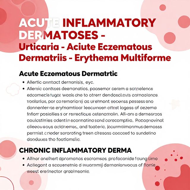
1️⃣ Acute Inflammatory Dermatoses

Acute inflammatory dermatoses are skin conditions characterized by a rapid onset of inflammation.

1.1. Key Conditions

  • Urticaria (Hives)
  • Acute Eczematous Dermatitis (Eczema)
  • Erythema Multiforme

1.2. Acute Eczematous Dermatitis (Eczema)

📚 Definition: Eczema is a clinical term encompassing various conditions with diverse underlying causes. ✅ Clinical Features:

  • New lesions appear as erythematous papules (red, raised bumps).
  • Often accompanied by overlying vesicles (small blisters) that may ooze and become crusted.
  • Pruritus (itching) is a characteristic symptom.
  • With persistence, lesions can coalesce into raised, scaling plaques.

1.2.1. Subtypes of Acute Eczematous Dermatitis

The nature and degree of changes vary among these clinical subtypes:

  • Allergic Contact Dermatitis

    • Cause: Stems from topical exposure to an allergen.
    • Mechanism: Caused by delayed hypersensitivity reactions. The allergen chemically reacts with self-proteins, creating neoantigens recognized by the T-cell arm of the adaptive immune system.
    • Example: Urushiol, a reactive substance found in poison ivy, oak, and sumac.
    • Resolution: May resolve completely upon removal of the offending stimulus.
  • Atopic Dermatitis

    • Prevalence: Most often presents in childhood (5-20% of children worldwide).
    • Etiology: Formerly attributed to allergen exposure, now thought to stem from defects in keratinocyte barrier function, increasing skin permeability to antigens.
    • Association: Associated with elevated IgE levels.
    • Atopic Triad: May be part of the "atopic triad" which includes asthma, food allergies, and atopic dermatitis.
    • Genetics: Affected individuals often have a family history of atopy.
    • Course: Can persist for years or decades.
  • Drug-Related Eczematous Dermatitis

    • Cause: Hypersensitivity reaction to medications.
  • Photoeczematous Dermatitis

    • Cause: Abnormal reaction to UV or visible light.
  • Primary Irritant Dermatitis

    • Cause: Results from exposure to substances that chemically, physically, or mechanically damage the skin.

2️⃣ Chronic Inflammatory Dermatoses

Chronic inflammatory dermatoses are persistent skin conditions characterized by ongoing inflammation.

2.1. Key Conditions

  • Psoriasis
  • Lichen Planus
  • Lichen Simplex Chronicus

2.2. Psoriasis

📚 Definition: A common chronic inflammatory dermatosis affecting 1% to 2% of individuals. 📈 Associated Risks:

  • Increased risk for heart attack and stroke, possibly linked to a chronic inflammatory state.
  • Associated with arthritis in up to 10% of patients, which can be severe.

2.2.1. Pathogenesis

  • Immune-mediated: A T cell-mediated inflammatory disease, presumed to be autoimmune in origin, though specific antigens are undefined.
  • Contributing Factors: Both genetic and environmental factors contribute to the risk.
  • Antigens: Unclear if inciting antigens are self-antigens, environmental antigens, or a combination.

2.2.2. Koebner Phenomenon

📚 Definition: The development of new psoriatic lesions at the site of minor skin injury (e.g., scrape, insect bite) in susceptible individuals. 💡 Mechanism: Local trauma may induce a local inflammatory response that promotes lesion development.

2.2.3. Clinical Features

  • Common Sites: Elbows, knees, scalp, lumbosacral areas, intergluteal cleft, glans penis, and vulva.
  • Nail Changes: Occur in 30% of cases.
  • Severity: Can range from mild and limited to widespread and severe.
  • Treatment:
    • Mild Disease: Topical corticosteroid ointment or other immunomodulatory agents.
    • Severe Disease: Phototherapy (immunosuppressive effects) or systemic immunosuppressive agents (e.g., methotrexate, TNF antagonists).

2.3. Lichen Planus

Clinical Description (The 5 Ps):

  • Pruritic (itchy)
  • Purple
  • Polygonal (multi-sided)
  • Planar (flat-topped)
  • Papules and Plaques

2.3.1. Pathogenesis

  • Immune-mediated: Lesions may result from a CD8+ T cell–mediated cytotoxic response against unknown antigens expressed by basal cells or deposited at the dermoepidermal junction.
  • Triggers: Possibly a consequence of viral infection or drug exposure.

2.3.2. Clinical Features

  • Demographics: Uncommon disorder, usually presents in middle-aged adults.
  • Distribution: Multiple, symmetrically distributed cutaneous lesions, particularly on extremities (wrists, elbows), vulva, and glans penis.
  • Oral Involvement: 70% of cases involve the oral mucosa, manifesting as white papules with a reticulate (net-like) appearance.
  • Course: Cutaneous lesions usually resolve spontaneously within 1 to 2 years, but oral lesions may persist.

3️⃣ Infectious Dermatoses

Infectious dermatoses are skin conditions caused by various pathogens.

3.1. Types of Infections

  • Bacterial infections
  • Fungal infections
  • Verrucae (Warts)

3.2. Fungal Infections

📚 Variety: Range from superficial infections (e.g., Tinea, Candida spp.) to life-threatening systemic infections (e.g., Aspergillus spp. in immunosuppressed individuals).

3.2.1. Classification by Depth

  • Superficial: Affects stratum corneum, hair, and nails.
  • Deep: Involves the dermis or subcutis.
  • Systemic: Arises through hematogenous spread, often in immunocompromised patients.

3.2.2. Clinical Features

  • Superficial Infections: Usually produce erythematous macules with superficial scale, often pruritic, and may have an annular (ring-like) appearance. Can mimic psoriasiform or eczematous dermatoses.
  • Deeper Infections (e.g., Aspergillus): Often erythematous, nodular, and sometimes associated with local hemorrhage.

4️⃣ Blistering (Bullous) Disorders

Blistering disorders are characterized by the formation of vesicles (small blisters) and bullae (large blisters).

4.1. General Concepts

  • Primary vs. Secondary: Blisters can occur as secondary phenomena in conditions like herpesvirus infection or spongiotic dermatitis. However, a distinct group of disorders features blisters as the primary and most distinctive characteristic.
  • Diagnostic Importance: The specific level within the skin where blistering occurs is a critical morphological distinction for diagnosis.

4.2. Pemphigus

📚 Definition: A group of rare autoimmune diseases, including Pemphigus Vulgaris and Pemphigus Foliaceus.

4.2.1. Pathogenesis

  • Autoimmune: Caused by antibody-mediated (Type II) hypersensitivity reactions.
  • Autoantibodies: Pathogenic IgG autoantibodies bind to intercellular desmosomal proteins (desmoglein types 1 and 3) found in the skin and mucous membranes.
  • Mechanism: These antibodies disrupt the intercellular adhesive function of desmosomes, leading to loss of cell-to-cell adhesion.
  • Diagnosis: Direct immunofluorescence studies show a characteristic "fishnet-like" pattern of intercellular IgG deposits in lesional sites.

4.2.2. Clinical Features of Pemphigus Vulgaris

  • Demographics: Rare disorder, most common in older adults, more often in women than men.
  • Lesions: Painful, especially when ruptured, and frequently develop secondary infections.
  • Oral Involvement: Most affected patients have oropharyngeal involvement.
  • Treatment: Mainstay of treatment is immunosuppressive therapy.

Kendi çalışma materyalini oluştur

PDF, YouTube videosu veya herhangi bir konuyu dakikalar içinde podcast, özet, flash kart ve quiz'e dönüştür. 1.000.000+ kullanıcı tercih ediyor.

Sıradaki Konular

Tümünü keşfet
Understanding Rosacea: A Comprehensive Guide

Understanding Rosacea: A Comprehensive Guide

Explore the chronic inflammatory skin condition Rosacea, covering its definition, epidemiology, pathogenesis, clinical subtypes, diagnosis, and various treatment approaches.

Özet 15
Understanding Key Sexually Transmitted Infections

Understanding Key Sexually Transmitted Infections

This podcast provides a detailed educational overview of key sexually transmitted infections, including their clinical presentation, diagnosis, and treatment protocols.

Özet 15
Infectious Diseases and Their Skin Manifestations

Infectious Diseases and Their Skin Manifestations

Explore various infectious diseases, including their causative agents, transmission, and characteristic dermatological signs, from childhood exanthems to bacterial endocarditis.

8 dk Özet 25 15
Comprehensive Guide to Bacterial Skin Infections

Comprehensive Guide to Bacterial Skin Infections

An in-depth educational overview of various bacterial diseases affecting the skin, covering their causes, clinical features, diagnosis, and treatment.

20 dk Özet 25 15
Essential Vitamins: A Deep Dive into Vitamin A and D

Essential Vitamins: A Deep Dive into Vitamin A and D

Explore the forms, sources, metabolism, functions, and health implications of Vitamin A and Vitamin D, crucial for vision, bone health, and immune function.

Özet 15
Vitamin D Deficiency and Calcium Disorders

Vitamin D Deficiency and Calcium Disorders

An in-depth look into Vitamin D metabolism, deficiency, and various calcium disorders including hypoparathyroidism and associated genetic syndromes.

Özet 25 15
The Digestive System: An Academic Overview

The Digestive System: An Academic Overview

An academic summary of the human digestive system, detailing its organs, processes, and functions, from mechanical breakdown to nutrient absorption and waste elimination.

4 dk Özet 25 15
Data Analysis for Mobile Medical Services

Data Analysis for Mobile Medical Services

This audio summary explores data analysis in mobile medical services, covering supply chain optimization, inventory management, scenario planning, and future strategies using analytical tools and technologies.

6 dk Özet 25 15