This study material has been compiled from a combination of copy-pasted text and an audio lecture transcript.
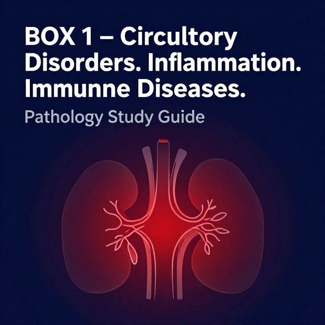
📚 Pathology Study Guide: Circulatory, Inflammatory, and Immune Disorders
Introduction
This study guide provides a comprehensive overview of various pathological conditions affecting different organ systems, focusing on their macroscopic (Gross Appearance - G/A) and microscopic (Microscopic Examination - M/E) features. Understanding these characteristics is crucial for diagnosing and comprehending disease processes in pathology.
1. Circulatory Disorders 🩸
1.1 Ischemic Renal Infarction
A common condition primarily caused by thromboemboli, often originating from the heart (e.g., mural thrombi in the left atrium, myocardial infarction, vegetative endocarditis, aortic aneurysm).
- G/A:
- Often multiple and may be bilateral.
- Pale or anemic, wedge-shaped lesions with the base resting under the capsule and the apex pointing towards the medulla.
- A narrow rim of preserved renal tissue under the capsule, spared due to blood supply from capsular vessels.
- M/E:
- Coagulative necrosis of renal parenchyma, appearing as "ghosts" of renal tubules and glomeruli without intact nuclei and cytoplasmic content.
- The margin of the infarct shows an inflammatory reaction, initially acute, later dominated by macrophages (MAPHs) and fibrous tissue.
1.2 Chronic Congestion of the Liver
Results from prolonged venous congestion, often due to heart failure.
- G/A:
- Liver is enlarged and tender, with a tense capsule.
- Cut surface exhibits a distinctive "nutmeg appearance" 🌰 – a red and yellow mottled pattern corresponding to congested central zones of lobules and fatty peripheral zones.
- M/E:
- Changes are most marked in the centrilobular zone (Zone 3), which is farthest from the blood supply and bears the brunt of hypoxia.
- Central veins and adjacent sinusoids are distended and filled with blood.
- Centrilobular hepatocytes show degenerative changes, leading to centrilobular hemorrhagic necrosis.
- Long-standing cases can develop fine centrilobular fibrosis and regeneration of hepatocytes, potentially leading to cardiac cirrhosis.
- Peripheral zones (less severely affected) may show fatty change in hepatocytes.
1.3 Pulmonary Edema
Accumulation of excess fluid in the lungs.
- G/A:
- Lungs are heavy, moist, and subcrepitant.
- Cut surface reveals frothy fluid (a mixture of air and fluid).
- M/E:
- Alveolar capillaries are congested.
- Initially, excess fluid collects in interstitial lung spaces in septal walls (interstitial edema).
- Later, fluid fills alveolar spaces (alveolar edema), forming brightly eosinophilic pink lines along the alveolar margin called hyaline membranes.
- Eosinophilic, granular, and pink proteinaceous material is present, often admixed with red blood cells (RBCs) and alveolar macrophages.
- Clinical Correlation: Fluid accumulates more in basal regions of the lungs. Chest X-ray (CXR) may show Kerley's lines, indicating thickened interlobular septa with dilated lymphatics.
1.4 Microhemorrhages of the Brain
Small hemorrhages within the brain, often associated with hypertension.
- M/E:
- A central core of clotted blood surrounded by a rim of brain tissue showing anoxic neuronal and glial changes and edema.
- Hyaline arteriosclerosis (pink amorphous matter) is typically seen in the vascular wall of the ruptured small vessel.
1.5 Thrombus Formation and Fate 📈
1.5.1 Mixed Thrombus
Thrombi vary in shape, size, and composition depending on their site of origin and blood flow rate.
- G/A:
- Arterial thrombi: White and mural.
- Venous thrombi: Red and occlusive.
- Mixed/laminated thrombi: Exhibit alternate white and red layers, known as "lines of Zahn."
- Red thrombi: Soft, red, and gelatinous.
- White thrombi: Firm and pale.
- M/E:
- Lines of Zahn are formed by alternate layers of light-staining aggregated platelets admixed with fibrin meshwork and dark-staining layers of red cells.
- Red (venous) thrombi contain more abundant red cells, leukocytes, and platelets entrapped in a fibrin meshwork, closely resembling blood clots in vitro.
1.5.2 Organizing and Recanalizing Thrombus
The fate of a thrombus involves either resolution or organization.
- Resolution:
- The thrombus activates the fibrinolytic system, releasing plasmin, which may dissolve the thrombus completely.
- Lysis is complete in small venous thrombi, but large thrombi may not be fully dissolved.
- Fibrinolytic activity can be accentuated by thrombolytic substances (e.g., urokinase, streptokinase), especially in the early stages when fibrin is in monomeric form (e.g., thrombolytic therapy for acute myocardial infarction).
- Organization:
- If the thrombus is not removed, it undergoes organization.
- 1️⃣ Phagocytic cells (neutrophils and macrophages) appear and phagocytose fibrin and cell debris.
- 2️⃣ Proteolytic enzymes liberated by leukocytes and endothelial cells begin digesting the coagulum.
- 3️⃣ Capillaries grow into the thrombus from the site of attachment, and fibroblasts invade, forming fibrovascular granulation tissue.
- 4️⃣ This tissue becomes dense and less vascular, eventually covered by endothelial cells.
- The thrombus is excluded from the vascular lumen and becomes part of the vessel wall.
- New vascular channels within it may re-establish blood flow ("recanalization").
- A fibrosed thrombus can undergo hyalinization and calcification (e.g., phleboliths in pelvic veins).
1.6 Hemorrhagic Pulmonary Infarction
Occurs due to embolism of pulmonary arteries.
- 💡 Note: The lungs receive a dual blood supply from both pulmonary and bronchial arteries. Therefore, occlusion of a pulmonary artery ordinarily does not produce infarcts unless the bronchial circulation is compromised or there is pre-existing lung disease.
- G/A:
- Wedge-shaped with the base on the pleura, hemorrhagic, variable in size, and mostly found in the lower lobes.
- Fibrinous pleuritis usually covers the area of infarct.
- Cut surface is dark purple, with a blocked vessel near the apex of the infarcted area.
- M/E:
- Coagulative necrosis of alveolar walls.
- Initially, infiltration by neutrophils and intense alveolar capillary congestion.
- Later, the area is replaced by hemosiderin-laden macrophages, phagocytes, and granulation tissue.
2. Inflammatory Conditions 🔥
2.1 Pneumonia
2.1.1 Focal Serous Pneumonia (Bronchopneumonia)
A patchy consolidation of the lungs, often centered around bronchioles.
- G/A:
- Patchy areas of red or grey consolidation, affecting one or more lobes, frequently bilateral, and more often involving lower zones due to gravitation of secretions.
- Cut surface is dry, granular, firm, red or grey, 3-4 cm in diameter, slightly elevated, and often centered around a bronchiole.
- M/E:
- Acute bronchiolitis.
- Suppurative exudate, chiefly neutrophils, in peribronchiolar alveoli.
- Thickening of alveolar septa by congested capillaries and leukocytic infiltration.
- Less involved alveoli contain edema fluid.
2.1.2 Lobar Fibrinous Pneumonia
A widespread consolidation of an entire lobe, progressing through four distinct stages:
- Stage of Congestion:
- Dilation and congestion of capillaries in the alveolar wall.
- Pale eosinophilic fluid in the air spaces, with few red cells and neutrophils.
- Bacteria present in the alveolar fluid.
- Red Hepatization:
- Marked extravasation of red cells and neutrophils, along with strands of fibrin.
- Many neutrophils have ingested bacteria.
- Alveolar septa are less prominent.
- Grey Hepatization:
- Dense fibrin strands.
- Reduced cellular exudate (fewer neutrophils and erythrocytes).
- Macrophages begin to appear.
- Resolution:
- Macrophages are predominant in the infiltrate.
- Granular and fragmented strands of fibrin are seen in alveolar spaces.
- Alveolar capillaries remain engorged.
2.2 Fibrinous Pericarditis
Inflammation of the pericardium, the fibroserous sac enclosing the heart.
- The pericardium is covered with a pink fibrinous exudate (pink-red amorphous matter on H&E stain).
- Pericardial Layers:
- Serous pericardium (innermost): Visceral layer (epicardium) and parietal layer.
- Fibrous pericardium (outermost).
- The mesothelium, a monolayer of specialized pavement-like cells, lines serous cavities and provides a slippery, non-adhesive, and protective surface.
2.3 Purulent Nephritis
Inflammation of the kidney characterized by pus formation. (No further details provided in source).
2.4 Granulation Tissue
A key component of wound healing and chronic inflammation.
- G/A:
- The floor of the lesion contains pink granulations composed of vascular connective tissue.
- Edges are sloping and bluish-white.
- M/E:
- Surface of the ulcer: Mixture of blood, fibrin, and inflammatory exudate.
- Zone underneath: Granulation tissue composed of proliferating fibroblasts, newly-formed small blood vessels, and varying numbers of inflammatory cells.
- Initially, polymorphs are present.
- Later stages: Macrophages and lymphocytes predominate.
- Epithelium grows from the edge of the wound as spurs.
- Granulation tissue matures from below upwards; in the late stage, it consists of dense collagen, scanty vascularity, and fewer inflammatory cells.
2.5 Acute Phlegmonous Appendicitis
The most common acute abdominal condition requiring surgical intervention.
- G/A:
- Appendix is swollen.
- Serosa is hyperemic and coated with fibrinopurulent exudate.
- Mucosa is ulcerated and sloughed.
- M/E:
- Neutrophilic infiltration of the muscularis propria.
- Mucosa is sloughed, and blood vessels in the wall are thrombosed.
- Periappendiceal inflammation is present in more severe cases.
3. Granulomatous Inflammation 🔬
3.1 "Foreign Body" Granuloma
A type of granuloma formed in response to inert foreign material.
- M/E:
- Presence of multinucleate giant cells (often "Foreign body" type, with centrally located nuclei).
- Lymphoid cells surrounding the lesion.
- The foreign body itself (e.g., filaments from surgical stitches) is often visible.
3.2 Tuberculous Granuloma
The hallmark lesion of tuberculosis, typically found in lymph nodes.
- M/E:
- Caseating necrosis: Foci of structureless, eosinophilic substance containing granular debris.
- Specific granulomatous inflammatory reaction consisting of epithelioid cells, with interspersed Langhans' giant cells (multinucleated giant cells with nuclei arranged in a horseshoe or peripheral pattern).
- A peripheral mantle of lymphocytes surrounds the granuloma.
3.3 Actinomycosis
A chronic bacterial infection characterized by granulomatous inflammation with central suppuration.
- M/E:
- 1️⃣ Inflammatory reaction: Granuloma with central suppuration, forming abscesses.
- 2️⃣ Periphery: Chronic inflammatory cells, giant cells, and fibroblasts.
- 3️⃣ Center of abscess: Bacterial colony, known as a "sulphur granule," which shows radiating filaments with hyaline, eosinophilic, club-like ends (representing secreted immunoglobulins).
- Bacterial Stains: Gram-positive filaments, non-acid-fast, and stain positively with Gomori's methenamine silver (GMS) staining.
3.4 Sarcoidosis of Lymph Node
A multisystem disease of unknown etiology, characterized by non-caseating granulomas.
- M/E:
- 1️⃣ Non-caseating sarcoid granuloma: Composed of epithelioid cells, Langhans' and foreign body giant cells, surrounded peripherally by fibroblasts.
- 2️⃣ Sarcoid granulomas are often described as "naked" 💡 because they lack a peripheral rim of lymphocytes or have a paucity of lymphocytes.
- 3️⃣ In late stages, the granuloma may be enclosed by hyalinized fibrous tissue or replaced by a hyalinized fibrous mass.
- 4️⃣ Giant cells in sarcoid granulomas may contain certain cytoplasmic inclusions:
- Asteroid bodies: Eosinophilic and stellate-shaped structures.
- Schaumann's bodies (conchoid bodies): Concentric laminations of calcium and iron salts, complexed with proteins.
- Birefringent cytoplasmic crystals.
4. Immune-Mediated & Other Conditions 🛡️
4.1 Allergic Nasal Polyp
Benign growths in the nasal passages, often associated with allergic inflammation.
- M/E:
- Loose edematous connective tissue containing some mucous glands.
- Inflammatory cells, including lymphocytes, plasma cells, and notably, eosinophils.
- Polyps are covered by respiratory epithelium, which may show squamous metaplasia.
4.2 Hashimoto's Thyroiditis
An autoimmune disease leading to chronic inflammation and destruction of the thyroid gland.
- G/A (Classic Form):
- Diffuse, symmetric, firm, and rubbery enlargement of the thyroid (may weigh 100-300g).
- Cut surface is fleshy with accentuation of normal lobulations, but the gland retains its normal shape.
- M/E (Classic Form):
- Extensive infiltration of the gland by lymphocytes, plasma cells, immunoblasts, and macrophages, often forming lymphoid follicles with germinal centers.
- Decreased number of thyroid follicles, which are generally atrophic and often devoid of colloid.
- Follicular epithelial cells transform into Hurthle cells (degenerated state):
- Abundant oxyphilic/eosinophilic and granular cytoplasm due to a large number of mitochondria.
- Contain large, bizarre nuclei.
- Slight fibrous thickening of septa separating thyroid lobules.
- G/A & M/E (Fibrosing Variant - less common):
- Firm, enlarged thyroid with compression of surrounding tissues.
- Considerable fibrous replacement of thyroid parenchyma with less prominent lymphoid infiltrate.
4.3 Myocardial Scar (Healing of Infarction)
The end result of myocardial infarction healing, typically occurring over about 6 weeks.
- G/A:
- The infarcted area is replaced by a thin, grey-white, hard, shrunken fibrous scar compared to the adjacent uninvolved grey-brown myocardium.
- M/E:
- Replacement of an irregular area of myocardium by dense fibrocollagenous tissue with foci of entrapped groups of myocardial fibers.
- Neighboring myocardial fibers show compensatory hypertrophy.
- The affected area of healed infarct (old granulation tissue) contains an infiltrate of some pigmented macrophages, lymphocytes, plasma cells, and a few capillary-sized blood vessels.
- Staining: Often visualized with special stains like Van Gieson, which highlights collagen (red).
4.4 Cirrhosis of the Liver
A diffuse disease characterized by disorganized lobular architecture and the formation of regenerative nodules, separated by irregular bands of fibrosis.
- G/A:
- Categorized by nodule size:
- Micronodular: Nodules <3 mm.
- Macronodular: Nodules >3 mm.
- Mixed: Both small and large nodules are seen.
- Cut surface shows grey-brown nodules separated from one another by grey-white fibrous septa.
- Categorized by nodule size:
- M/E:
- The lobular architecture of hepatic parenchyma is lost, and central veins are hard to find.
- Hepatocytes in the surviving parenchyma form disorganized masses of hepatocytes, known as regenerative nodules.
- Fibrous septa contain some mononuclear inflammatory cell infiltrate and proliferated bile ductules.