This study material has been compiled and organized from a lecture audio transcript and supplementary text provided by the user.
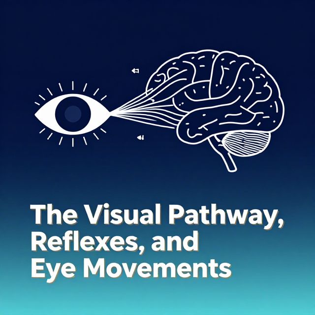
📚 Introduction to Visual Pathways
This study guide provides a comprehensive overview of the visual pathways, from the initial detection of light in the retina to complex processing in the cerebral cortex. It also details the visual defects associated with lesions at various points along these pathways and explores higher-order visual processing.
🎯 Learning Outcomes
Upon completing this study material, you should be able to:
- ✅ Describe the visual pathway from the retina to the primary visual cortex (V1).
- ✅ Explain the visual defects (e.g., anopias or anopsias) associated with lesions along this pathway.
- ✅ Briefly describe the function of the pathways extending from V1 to other cortical regions.
👁️ The Visual Pathway from Retina to Primary Visual Cortex (V1)
Vision is a complex process that begins with light entering the eye and culminates in the brain's interpretation of visual information.
1️⃣ Initial Vision Formation
- Light passes through the cornea and pupil.
- It is then focused by the lens to form a visual image on the retina.
- Initial detection occurs via specialized cells in the retina.
- Primary processing also begins within the retina itself.
- The output from the retina consists of axons from the retinal ganglion cells, which collectively form the optic nerve (Cranial Nerve II).
2️⃣ Key Elements of the Pathway to V1
The visual information travels through a precise sequence of structures:
- Optic Nerve: Carries signals from each eye.
- Optic Chiasm: A crucial point where partial decussation (crossing over) of nerve fibers occurs.
- Optic Tracts: Carry fibers from the chiasm.
- Lateral Geniculate Nucleus (LGN): Located in the thalamus, this is where the first synapse in the central visual pathway occurs, relaying information.
- Optic Radiation: Fibers extending from the LGN.
- Primary Visual Cortex (V1, Area 17): Located in the occipital lobe, this is where the optic radiation fibers synapse, marking the initial cortical processing of visual input.
3️⃣ Retinal Image Inversion and Hemifield Representation
💡 An important concept to grasp is how the visual field is mapped onto the retina and subsequently processed:
- The image of an object formed on the retina is inverted vertically and reversed horizontally.
- Partial Decussation at the Optic Chiasm:
- Information from the right temporal hemi-retina (outer half) remains ipsilateral (same side), traveling to the right LGN and then to the right V1.
- Information from the left nasal hemi-retina (inner half) crosses over at the optic chiasm to the right LGN and then to the right V1.
- This means the left side of the visual field is ultimately represented in the right LGN and right visual cortex, and vice versa for the right visual field.
4️⃣ Optic Radiation and Meyer's Loop
The optic radiation, carrying signals from the LGN to V1, has a specific anatomical organization:
- Lower Fibers (Meyer's Loop): Fibers originating from the lower half of the retina (representing the upper visual field) pass over the temporal horn of the lateral ventricle, forming Meyer's loop. These fibers enter V1 below the calcarine sulcus.
- ⚠️ Clinical Significance: Damage to Meyer's loop will cause defects in the upper visual field on the side opposite the lesion.
- Upper Fibers: Fibers originating from the upper half of the retina (representing the lower visual field) pass through the parietal lobe and enter V1 above the calcarine sulcus.
- Remember, due to retinal inversion, the top half of the visual image is processed by the lower half of the retina, and vice versa.
📊 Visual Defects from Lesions Along the Pathway to V1
Understanding the specific patterns of vision loss resulting from lesions at different points in the visual pathway is crucial for diagnosis.
- Optic Nerve (Pre-Chiasm Lesion):
- Defect: Ipsilateral monocular blindness.
- Explanation: Complete loss of vision in one eye, affecting both left and right visual fields of that single eye.
- Optic Chiasm Lesion:
- Defect: Bi-temporal hemianopia.
- Explanation: Loss of the temporal (outer) visual fields in both eyes, often described as "tunnel vision." This occurs because the crossing nasal fibers are affected.
- Optic Tract (Post-Chiasm, Pre-LGN Lesion):
- Defect: Contralateral homonymous hemianopia.
- Explanation: Loss of the same side of the visual field in both eyes (e.g., the entire left half of vision in both eyes). This is because the optic tract carries information from the contralateral visual field.
- Whole Optic Radiation or Primary Visual Cortex (V1) Lesion:
- Defect: Contralateral homonymous hemianopia.
- Explanation: Similar to an optic tract lesion, resulting in loss of the same side of the visual field in both eyes.
- Meyer's Loop Lesion:
- Defect: Contralateral superior quadrantanopia.
- Explanation: Loss of the upper quadrant of vision on the side opposite the lesion (e.g., a lesion in the right Meyer's loop causes loss of the upper left visual field in both eyes).
🧠 Visual Pathways After V1 and Higher-Order Processing
While V1 is essential for initial visual processing, it is not sufficient for complete visual perception.
1️⃣ Beyond V1: Cortical Blindness and Associated Areas
- Loss of V1 can lead to cortical blindness, meaning the eyes and optic nerves may be intact, but the brain cannot process visual input.
- However, V1 alone is not enough for full cortical processing. A network of associated cortical areas (V2-V5) receives information from V1, further processing and relaying signals.
- Riddoch's Phenomenon/Syndrome: Interestingly, some patients with V1 lesions and cortical blindness can still respond to moving objects, suggesting alternative or residual pathways for motion detection.
2️⃣ Two Main Pathways for Higher-Level Processing
From V1, visual information diverges into two primary streams:
a. 📈 The Dorsal (Parietal) Pathway: "Where" or "How" Pathway
- Origin: Primarily derives input from the magnocellular portion of the LGN.
- Function: Crucial for processing information related to movement, spatial awareness, and guiding actions. It helps us understand "where" objects are and "how" to interact with them.
- Lesions: Can result in akinetopsia, the loss of perception of movement.
- Example (Patient LM): A patient with akinetopsia struggled with pouring liquids (appeared frozen), felt insecure in rooms with moving people (they "suddenly appeared"), and couldn't cross streets due to inability to judge car speed, despite being able to identify objects.
b. 🎨 The Ventral (Temporal) Pathway: "What" Pathway
- Origin: Primarily derives input from the parvocellular portion of the LGN.
- Function: Primarily involved in processing color, form, and object recognition. It helps us understand "what" an object is.
- Lesions: Can lead to visual agnosia, an inability to identify objects by sight, even though the person can see them and may perceive movement.
- Specialized Forms: Visual agnosia can be highly specific. For example, prosopagnosia is the inability to recognize faces, even familiar ones, while still knowing it is a face.
- Recommended Reading: "The Man Who Mistook His Wife for a Hat" by Oliver Sacks provides vivid examples of such conditions.
📝 Summary
The visual system is an intricate network, and understanding its components is key to appreciating visual perception and the impact of neurological damage.
- The pathway from the retina to V1 involves a precise sequence of structures, including the optic nerve, chiasm, tracts, LGN, and optic radiation.
- Lesions at different points along this pathway result in distinct and predictable patterns of visual field loss.
- Beyond V1, visual information is processed by two main pathways: the dorsal (parietal) pathway for movement and spatial awareness, and the ventral (temporal) pathway for object recognition and form.
- Damage to these higher pathways can lead to conditions like akinetopsia and visual agnosia, profoundly affecting how individuals perceive and interact with their visual world.