Source Information: This study material has been compiled from copy-pasted text and a lecture audio transcript.
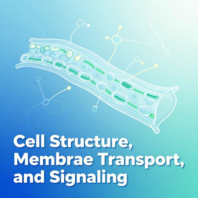
📚 Comprehensive Guide to Cell Structure, Membrane Transport, and Signaling
Introduction
Cells are the fundamental units of life, performing all essential biological functions. Understanding their basic components, intricate internal structures, mechanisms for transporting substances, and methods of communication is crucial for comprehending biological processes. This guide provides a structured overview of these core cellular concepts.
I. Basic Cell Components & Eukaryotic Structure
All cells share fundamental components necessary for life. ✅ Basic Cell Parts:
- Cell Membrane: Surrounds the cell, controlling the movement of substances in and out.
- Cytoplasm: The fluid interior where organelles are located and cellular reactions occur.
- Genetic Material: Contains hereditary information and controls cell activities.
💡 Prokaryotic vs. Eukaryotic Cells: Both types possess the cell membrane, cytoplasm, and genetic material. However, eukaryotic cells are distinguished by the presence of a nucleus, which encloses their genetic material.
Eukaryotic cells exhibit a complex internal organization with various specialized organelles:
- Nucleus
- Cytoplasm
- Ribosomes
- Endoplasmic Reticulum (ER)
- Golgi Apparatus
- Mitochondria
- Lysosomes
- Peroxisomes
II. The Cytoskeleton & Cell Junctions
A. The Cytoskeleton
The cytoskeleton is a dynamic network of protein fibers within the cytoplasm that provides structural support, facilitates movement, and aids in intracellular transport. ✅ Main Components:
- Microfilaments:
- Smallest fibers, made of actin protein.
- Involved in cell movement (e.g., muscle contraction, cell crawling).
- Support the cell membrane and maintain cell shape.
- Crucial for cytokinesis (cell division).
- Microtubules:
- Largest fibers, made of tubulin protein.
- Maintain cell shape and provide structural support.
- Form the mitotic spindle during cell division.
- Serve as tracks for intracellular transport of vesicles and organelles.
- Form structures like cilia and flagella.
- Intermediate Filaments: Provide mechanical strength and help anchor organelles.
B. Cell Junctions
Cell junctions are specialized structures that connect neighboring cells, maintaining tissue integrity, enabling communication, and regulating substance movement.
- Tight Junctions:
- Function: Form an impermeable seal between adjacent cells, preventing substances from leaking between them. They also maintain cell polarity.
- Analogy: 🗄️ Zipper
- Example: Commonly found in epithelial tissues (e.g., intestine, kidney).
- Desmosomes:
- Function: Strong anchoring junctions that hold cells together, providing mechanical strength by connecting intermediate filaments. They reduce the chance of cells tearing under stress.
- Analogy: 🔗 Velcro
- Example: Important in tissues under stress (e.g., skin, heart muscle).
- Gap Junctions:
- Function: Communication channels that allow small molecules and ions to pass directly between cells, enabling electrical and chemical signaling.
- Analogy: 🚇 Tunnel
- Example: Essential in heart and smooth muscle tissue, allowing rapid spread of electrical signals for synchronized contraction (e.g., in cardiac muscle cells, they enable the heart to beat as a functional syncytium).
III. The Cell Membrane: Structure & Fluidity
A. Importance of the Cell Membrane
The cell membrane (plasma membrane) is a thin, flexible, and selectively permeable layer that surrounds the cell. ✅ Key Roles:
- Controls what enters and leaves the cell.
- Maintains internal cellular balance (homeostasis).
- Facilitates communication with the environment.
- Involved in nutrient uptake and waste removal.
- Plays a role in drug and anesthetic interactions.
- Critical for cell signaling, inflammation, and healing.
- Generates and transmits electrical impulses in nerves and muscles.
- Regulates cell growth and proliferation.
📚 Cell Growth vs. Proliferation:
- Cell Growth: Refers to an increase in the total mass of a cell (cytoplasmic, nuclear, and organelle volume increase). Happens before proliferation.
- Cell Proliferation: The process by which a cell grows and then divides to produce two daughter cells.
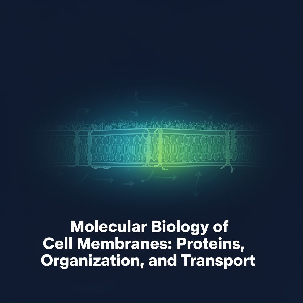
B. Structure of the Cell Membrane (Fluid Mosaic Model)
The cell membrane is a fluid, dynamic structure primarily composed of a phospholipid bilayer. ✅ Main Components:
- Phospholipid Bilayer: Forms the basic structure. Phospholipids have a hydrophilic (water-loving) head pointing outwards and hydrophobic (water-fearing) tails pointing inwards.
- Cholesterol: Stabilizes the membrane and acts as a fluidity buffer.
- Membrane Proteins: Act as channels, carriers, receptors, enzymes, or anchoring points.
- Carbohydrates: Help in cell recognition and adhesion.
C. Phospholipids and Membrane Fluidity
The fluidity of the cell membrane is crucial for its function and is significantly influenced by the type of fatty acid tails in its phospholipids. 📊 Saturated vs. Unsaturated Lipids:
| Feature | Saturated Lipids | Unsaturated Lipids | | :--------------------- | :--------------------------------------------- | :------------------------------------------------- | | Carbon–carbon bonds | Only single bonds | One or more double bonds | | Hydrogen content | Fully saturated with hydrogen | Not fully saturated with hydrogen | | Molecular shape | Straight chains | Bent (kinked) chains | | Physical state (room temp.) | Usually solid | Usually liquid | | Membrane effect | Make membranes less fluid, more rigid | Increase membrane fluidity | | Common sources | Butter, animal fat | Olive oil, sunflower oil, fish oil |
💡 Why it Matters: Saturated lipids pack tightly, leading to a more rigid membrane. Unsaturated lipids, with their "kinks," pack loosely, increasing membrane flexibility and fluidity. This fluidity is essential for transport, receptor function, and cell signaling.
D. Cholesterol's Role
Cholesterol regulates membrane fluidity, preventing it from becoming too rigid at low temperatures or too fluid at high temperatures. However, excessive cholesterol accumulation can increase membrane rigidity and decrease permeability, potentially leading to fragility.
E. Carbohydrates
Found on the outer surface, attached to proteins (glycoproteins) or lipids (glycolipids). Together, they form the glycocalyx, a protective and recognition layer. ✅ Functions: Cell recognition, cell adhesion, immune recognition, cell communication.
F. Membrane Proteins
Membrane proteins are diverse and perform various critical functions:
- Transport: Move substances across the membrane (channels, carriers, pumps).
- Receptors: Receive signals from outside the cell.
- Enzymes: Catalyze chemical reactions.
- Anchoring: Connect the cell to other cells or the cytoskeleton. Proteins can be integral (pass through the membrane) or peripheral (attach to one side).
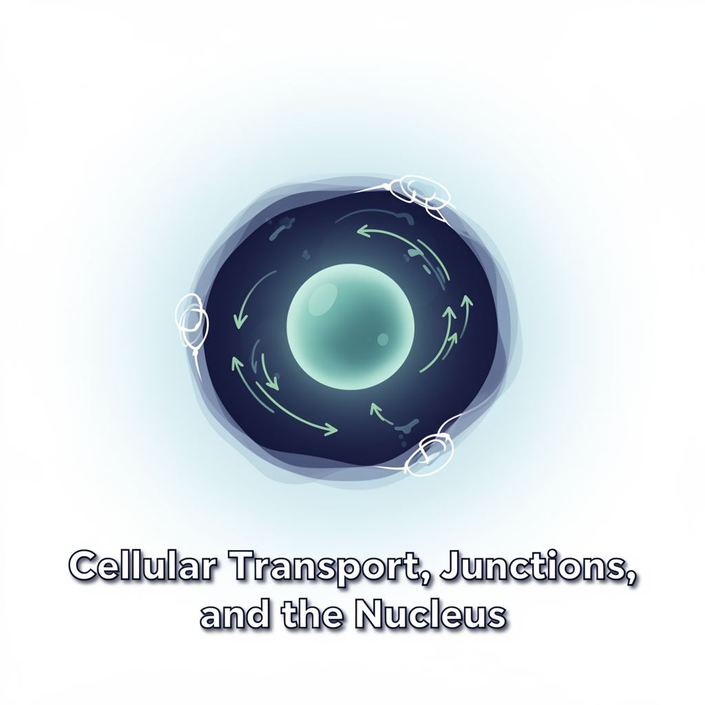
IV. Membrane Transport Mechanisms
The cell membrane's selective permeability allows it to control the movement of substances.
A. Passive Transport (No Energy Required)
Moves substances from a region of high concentration to low concentration.
- Simple Diffusion: Small, lipid-soluble molecules (e.g., O₂, CO₂) pass directly through the membrane.
- Facilitated Diffusion: Transport proteins (channels or carriers) help larger or charged molecules (e.g., glucose, ions) cross the membrane.
- Osmosis: The movement of water across a semi-permeable membrane from an area of low solute concentration (more water) to high solute concentration (less water).
- Hypotonic solution: Water enters the cell → cell swells.
- Hypertonic solution: Water leaves the cell → cell shrinks.
- Isotonic solution: No net water movement → cell size stays the same.
B. Active Transport (Energy Required)
Moves substances against their concentration gradient (from low to high concentration) and requires ATP energy.
- Example: The Sodium-Potassium pump (Na⁺/K⁺ pump) maintains ion balance, crucial for nerve transmission and muscle function.
C. Vesicular Transport
Transports large materials using membrane-bound vesicles.
- Endocytosis: The cell takes substances inside.
- Pinocytosis: "Cell drinking" – uptake of fluids and dissolved solutes.
- Phagocytosis: "Cell eating" – uptake of large particles or microorganisms.
- Receptor-mediated endocytosis: Specific uptake of molecules that bind to receptors.
- Exocytosis: The cell releases substances outside (e.g., secretion of hormones, enzymes).
V. Cell Signaling: Information Transmission
Cells communicate constantly using signaling molecules that bind to membrane receptors, triggering specific responses. Signals can originate from distant locations (endocrine), nearby cells (paracrine), or the same cell (autocrine).
3️⃣ Stages of Cell Signaling:
- Reception:
- A signaling molecule (ligand) binds to a specific receptor protein.
- Membrane Receptors: Located on the cell surface (e.g., G protein-coupled receptors, receptor tyrosine kinases).
- Intracellular Receptors: Located inside the cell (cytoplasm or nucleus) for small or hydrophobic ligands (e.g., steroid hormones).
- Transduction:
- Binding of the ligand causes a change in the receptor's shape, initiating a signal relay inside the cell.
- Often involves multi-step pathways, amplifying the signal.
- Phosphorylation Cascades: Protein kinases add phosphate groups (phosphorylation) to activate proteins, while protein phosphatases remove them (dephosphorylation) to deactivate.
- Second Messengers: Small, non-protein molecules or ions (e.g., cAMP, Ca²⁺) relay signals within the cell.
- Response:
- The transduced signal triggers a specific cellular activity.
- Examples: Regulation of gene expression (turning genes on/off), changes in protein activity (e.g., opening ion channels), altered cell metabolism, or major cellular events like cell division or apoptosis (programmed cell death).
VI. Clinical Relevance in Dentistry
The principles of cell structure, membrane transport, and signaling are highly relevant to oral health and dental treatments.
- 💊 Drug and Anesthetic Effects: Understanding how drugs interact with cell membranes and receptors is crucial for effective pain management and treatment.
- 🩹 Oral Tissue Healing: Cell signaling pathways regulate inflammation and tissue repair after injury or surgery.
- 🛡️ Immune Responses: Cellular communication is vital for the immune system to combat infections in the oral cavity.
- 💧 Saliva Secretion: Membrane transport mechanisms are essential for the proper function of salivary glands.
- ⚡ Pain Transmission: Ion channels and electrical impulses across nerve cell membranes are fundamental to how dental pain is perceived and transmitted.
Conclusion
The cell membrane is a dynamic and critical structure that controls substance entry and exit. Passive transport occurs without energy, while active transport requires ATP. Vesicular transport handles large materials. Furthermore, membrane receptors facilitate vital cellular communication through reception, transduction, and response. These intricate cellular mechanisms are fundamental to overall health and hold particular importance in various dental treatments and oral health processes.