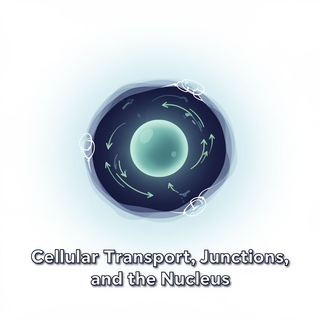
Cellular Transport, Junctions, and the Nucleus: A Comprehensive Study Guide
Source Information: This study material has been compiled from a copy-pasted text and a lecture audio transcript.
📚 Introduction to Cellular Dynamics
Cells are dynamic entities, constantly interacting with their environment and maintaining internal organization. This study guide explores the intricate mechanisms of macromolecule transport across membranes, the specialized structures that connect cells, and the central role of the nucleus in cellular function and heredity. Understanding these processes is fundamental to comprehending cellular physiology and pathology.
1. 🚚 Macromolecule Transport
Macromolecules, such as proteins, nucleic acids, and complex carbohydrates, move within and between cells through various mechanisms. While vesicular transport is a primary route, several direct transport pathways also exist.
1.1. Direct Transport of Macromolecules
Some macromolecules bypass vesicular transport and move directly across membranes. ✅
- Nuclear Pores: Transport of molecules between the nucleus and cytoplasm.
- Mitochondrial Import: Cytosolic proteins are imported directly into mitochondria.
- Fusion Pores: Elimination of secretion products at the cell periphery.
- Bacterial Conjugation: Direct transfer of nucleic acids (a primitive form of sexual reproduction).
- Viral Infection: Transfer of viral nucleic acids into host cells.
- Genetic Transformation: Experimental transfer of nucleic acids across the plasma membrane.
1.2. Vesicular Transport 📦
Eukaryotic cells extensively use vesicles for transporting materials across membranes or between organelles. This dynamic process is categorized into three main types:
- Exocytosis: Moving materials out of the cell.
- Endocytosis: Bringing materials into the cell.
- Transcytosis: Transcellular transport of materials.
2. 📤 Exocytosis
Exocytosis is the process by which cells eliminate various materials to the extracellular space.
- Mechanism: Macromolecules are packaged into vesicles, which are then moved along microtubules by cellular motors towards the plasmalemma. Upon reaching the cell periphery, these vesicles fuse with the plasma membrane, releasing their contents outside.
- Examples:
- Evacuation of cellular secretion products (e.g., hormones, enzymes).
- Release of neurotransmitters into the synaptic space.
- Expulsion of melanin granules from skin melanocytes, which are then phagocytosed by keratinocytes for skin pigmentation.
- Removal of undigested residues from lysosomes (cellular defecation).
- Release of lysosomal enzymes under physiological (e.g., osteoclasts) or pathological conditions (e.g., cellular regurgitation, shock).
3. 📥 Endocytosis
Endocytosis is the process of transporting materials from the extracellular space into the cell via vesicles. It is broadly classified based on the nature of the ingested material.
3.1. Phagocytosis (Cell Eating) 🍔
Phagocytosis involves the transport of solid materials into the cell.
- Role:
- Nutrition: A mode of nutrition in protozoa.
- Defense: In mammals and humans, specialized phagocytes play a crucial role in the immune system, capturing and destroying bacteria, parasites, foreign substances, cellular residues, and old/damaged cells.
- Phagocytes: Macrophages and neutrophilic leukocytes are key phagocytes, originating from bone marrow stem cells. Macrophages are also known as histiocytes. They form a phagocytic system with mobile (e.g., neutrophils in blood) and sessile components (e.g., macrophages in spleen, liver Kupffer cells).
- Quantitative Importance: Removal of old cells (e.g., 10^11 old erythrocytes daily by macrophages) is quantitatively more significant than destroying bacteria, though both are vital. Phagocytosis is the final step in fighting infections, as antibiotics primarily prevent bacterial multiplication.
- Steps of Phagocytosis:
- Chemotaxis: 🧭 Oriented movement of phagocytes towards an infection site, guided by signals (bacterial components, endogenous compounds like complement system components, lymphokines).
- Recognition and Attachment: 🤝 Phagocyte membrane receptors bind to particles (ligands). Old/malignant cells are recognized by sugar groups on their membranes (e.g., sialic acid removal from erythrocytes leads to rapid phagocytosis). Bacteria often require opsonization (coating with antibodies) before attachment.
- Engulfment (Inclusion): 🖐️ Phagocyte emits pseudopodia to surround the particle, enclosing it in a vesicle called a phagosome.
- Killing and Digestion: 🔪 Phagosome fuses with a late endosome or lysosome. An oxidase from the plasmalemma produces super-oxide anion (O2-), which converts to hydrogen peroxide (H2O2). H2O2 and O2- generate highly reactive free radicals (HO•) and singlet oxygen, effectively killing microbes.
- ⚠️ Clinical Relevance: Absence of this oxidase leads to Chronic Granulomatous Disease, a genetic condition where leukocytes cannot destroy most bacteria, resulting in persistent infections.
3.2. Pinocytosis (Cell Drinking) 💧
Pinocytosis involves the transport of liquid materials (macromolecules in solution) into the cell.
- Types:
- Receptor-Independent Pinocytosis (Fluid-Phase/Constitutive):
- Common in many cells (e.g., macrophages ingest equivalent of entire plasmalemma in 30 min).
- Introduces large amounts of macromolecules in solution.
- Cell surface and volume remain constant due to simultaneous addition of membrane material from the Golgi apparatus.
- Receptor-Mediated Pinocytosis:
- More selective, involving membrane receptors that recognize specific extracellular macromolecules.
- Mechanism: Ligand-receptor complexes diffuse into specialized regions called caveolae. These depressions are coated on their cytoplasmic face by a network of clathrin protein. Clathrin molecules form triskelions (three-footed structures) that polymerize into pentagonal or hexagonal networks. Caveolae then bud off to form vesicles, which subsequently lose their clathrin coat.
- Key Difference: Concentrates specific ligands within endosomes, unlike receptor-independent pinocytosis where concentrations mirror the extracellular fluid.
- Example: Cholesterol Capture (LDL) 📈
- Most cholesterol is transported in blood as Low-Density Lipoproteins (LDL).
- Cells needing cholesterol produce LDL receptors, which bind LDL with high specificity.
- LDL-receptor complexes move into clathrin-coated caveolae, forming vesicles.
- Vesicles lose clathrin, fuse with early endosomes.
- LDL dissociates from receptors; receptors are recycled back to the plasmalemma.
- Early endosome fuses with late endosome/lysosome to hydrolyze cholesterol esters.
- Free cholesterol enters cytosol, inhibiting new LDL receptor synthesis and cellular cholesterol synthesis.
- ⚠️ Clinical Relevance: Genetic defects in LDL receptors lead to severe hyper-cholesterolemia, premature atherosclerosis, and myocardial infarcts. Understanding this led to the development of statins (cholesterol synthesis inhibitors).
- Receptor-Independent Pinocytosis (Fluid-Phase/Constitutive):
4. 🔄 Transcytosis
Transcytosis is a trans-cellular transport mechanism where materials, enclosed in vesicles, pass unmodified through a cell to be released at the opposite pole, without being intercepted by lysosomes.
- Mechanism: Vesicles containing materials cross the cell from one side to the other.
- Historical Context: George Palade (1953) described vesicles crossing endothelial cells, suggesting their role in macromolecule exchange. Ion Baciu demonstrated its high speed. Nicolae and Maya Simionescu, with Palade, showed vesicles could move independently or form continuous channels.
- Example: Transit of antibodies from mother's milk through newborn enterocytes, conferring infection resistance.
5. 🔗 Cell Junctions
Cell junctions are stable, permanent structures of the plasmalemma specialized for intercellular communication and adhesion. There are three primary types:
5.1. Adherens Junctions (Desmosomes) 💪
These junctions keep cells together, providing mechanical strength to tissues under stress (e.g., epithelia, heart muscle, uterine cervix).
- Types:
- Belt Desmosomes (Zonula Adherens): Continuous ring of actin filaments at the apical end of epithelial cells, interconnected by integral adhesion proteins.
- Spot Desmosomes: Two dense cytoplasmic plaques under adjacent plasma membranes, interconnected by integral proteins. Anchor sites for keratin filaments (tonofilaments).
- Hemidesmosomes: Half the structure of spot desmosomes, attaching the cytoplasmic plaque to the basal lamina.
5.2. Tight Junctions 🔒
Regions where the intercellular space disappears, with adjacent membranes connected by junction proteins arranged in rows.
- Function: Prevent fluid leakage between cells, ensuring all substances cross the plasma membrane for absorption. They also maintain the distribution of transporters and membrane domains.
- Examples:
- Intestinal Epithelium: Glucose is actively transported at the apical side and passively transported into blood at the baso-lateral side.
- Blood-Brain Barrier: Formed by tight junctions between endothelial cells in brain blood vessels, protecting the brain from toxins while allowing essential substances (glucose, O2, CO2) to pass. Some toxins (ethanol, morphine) can bypass it. Proteins and most antibiotics cannot cross, but myelin can in newborns.
5.3. Communication Junctions (Gap Junctions & Synapses) 🗣️
Facilitate direct communication between cells.
- Gap Junctions:
- Most common in animal cells.
- Communication via protein structures called connexons, which cross the plasmalemma. Each connexon has 6 protein subunits (connexins).
- Form tube-shaped channels isolated from extracellular fluid, allowing direct cell-to-cell communication.
- Permeable to ions and small molecules (<1,000-1,500 Da) like glucides, amino acids, nucleotides, hormones, vitamins.
- Damage Response: If a cell is damaged, Ca2+ influx causes neighboring gap junctions to close, preventing lesion spread.
- Electrical Synapses:
- Essentially gap junctions that allow nervous impulses to pass without intermediates.
- 10,000 times more permeable to metallic ions than the rest of the membrane.
- Responsible for electrical coupling in myocardium and intestinal smooth muscle cells. These channels are dynamic.
6. 🚨 Implications of Membranes in Pathology
Changes at the membrane level are observed in virtually every disease.
- a) Genetic Diseases (Single Cell Type): Defects in specific membrane proteins (e.g., erythrocyte membrane protein defects leading to hemolytic anemias like spherocytosis, elliptocytosis).
- b) Genetic Diseases (Generalized Membrane Defects): Proteins in various cell types are affected, but disease manifests where proteins have crucial roles.
- Muscular Dystrophies: Absence/modification of dystrophin (muscle cell membrane protein) increases plasma membrane permeability, impairing muscle function.
- Channelopathies: Modified ionic transporters, primarily affecting the nervous system (e.g., genetic epilepsies) due to high impact on nerve cell properties.
- c) Genetic Diseases of Cell Organelles: Defects in organelle membrane proteins.
- d) Infectious Diseases:
- Viruses introduce genetic material through plasma membranes.
- Bacteria/parasites evade membrane-related defense systems (e.g., phagocytosis).
- Antibodies target pathogenic agent membranes.
- e) Autoimmune Diseases: Modified membrane structures are recognized as foreign, triggering immune responses (e.g., anemias, hepatitis).
- f) Cardiovascular Diseases: Involvement of ion channels in arterial hypertension, and LDL receptor defects in atheromatosis.
- g) Cancer: Changes in plasma membrane and intracellular membrane structures in malignant cells.
- h) Drug Interactions: Most drugs act by binding to membrane receptors or passing through cell membranes.
7. 🧠 The Cell Nucleus
The nucleus is the central command center of eukaryotic cells, performing two fundamental functions.
7.1. Roles and Components
- 1️⃣ Genetic Material Storage: Stores most of the cell's genetic material in chromosomes.
- 2️⃣ Cellular Coordination: Coordinates all cellular functions.
- Structural Variation:
- Interphase (Metabolic Nucleus): Contains nuclear envelope, chromatin, nucleolus, and nuclear matrix.
- Cell Division (Genetic Nucleus): Nuclear envelope disassembles, chromatin condenses into chromosomes.
7.2. Structure of Nucleus in Interphase
7.2.1. Nucleus in the Living Cell
- Study Techniques: Phase contrast or interferential contrast microscopy.
- Observations: Nuclear envelope at periphery, granular chromatin, dense nucleolus (up to 80% dry substance).
- Vital Role: Perforating the nuclear envelope or removing the nucleus kills the cell; perforating only the plasmalemma allows survival.
- Dynamics: Video-microscopy shows nuclear movements (rotation, translation, expulsion of degenerated nuclei).
- Biochemical Study: Differential centrifugation separates nuclei (denser, more alkaline pH than cytoplasm) for physical-chemical analysis.
7.2.2. Morphology and Ultrastructure (after fixation)
- Position: Usually central in young, spherical cells. Peripheral in differentiated cells (adipose, striated muscle) or near plasma membrane (secretory cells).
- Number: Typically unique. Some cells are binucleated (hepatocytes, chondrocytes). Osteoclasts can have up to 100 nuclei. Syncytial cells (e.g., striated muscle fibers) have hundreds.
- Shape: Generally corresponds to cell shape (spherical, ovoid, spindle, flat). Polymorphonuclear leukocytes have multi-lobed nuclei. Malignant cells often have aberrant shapes.
- Size: Average 5-12 µm (4 µm in sperm, 25 µm in oocyte).
- Nucleo-plasmatic Ratio: Ratio of nuclear volume to cytoplasmic volume (1/3 - 1/20). High in young cells with intense metabolism and malignant cells (around 1/3). Smaller in adult/old cells.
7.2.3. Nuclear Envelope 🛡️
A critical component with two membranes and pores.
- Outer Nuclear Membrane: Continuous with the rough endoplasmic reticulum (RER) membrane, may have ribosomes.
- Inner Nuclear Membrane: Adheres to nuclear content.
- Perinuclear Space: Lies between the two membranes, continuous with RER lumen.
- Nuclear Pores: Where the two membranes merge.
- Nuclear Lamina:
- Network of fibrous proteins under the inner nuclear membrane, between pores.
- Composed of intermediate filaments (lamins A, B, C).
- Functions: Maintains nuclear shape, organizes chromatin in interphase, crucial for nuclear envelope disassembly (prometaphase) and reassembly (telophase) during cell division.
- ⚠️ Clinical Relevance: A point mutation in the LMNA gene (chromosome 1) causes Hutchinson-Gilford Syndrome (Progeria), characterized by premature aging.
- Pores (Pore Complexes or Porosomes): Complex structures regulating transport between nucleus and cytoplasm.