This study material has been compiled from lecture slides (PDF/PowerPoint text) and an audio transcript provided by Prof. Dr. Elif Aylin Özüdoğru, Spring 2026, İstanbul.
Cell Division and Cell Cycle - I 🧬
1. Introduction to Cell Division
All living organisms, from the simplest bacteria to complex multicellular creatures, undergo fundamental processes such as growth, development, tissue regeneration, and reproduction. These processes are critically dependent on cell division ✅. Cell division enables organisms to produce offspring and transmit their hereditary information. The initial observation of cell division, specifically mitosis, was made by German embryologist Walther Fleming in 1882, who observed thread-like structures dividing within salamander larvae cells.
2. Prokaryotic Cell Division: Binary Fission 🦠
Prokaryotic cells, which lack a nucleus, divide through a simpler process called binary fission.
2.1. Process Overview
- The cell divides into two equal or nearly equal halves.
- Prokaryotic cells grow by increasing in cell number rather than individual cell size.
- Bacterial populations increase exponentially, doubling with each generation time.
2.2. Genetic Material and Replication
📚 Genome: The genetic information exists as a single, circular, double-stranded DNA molecule.
- Replication Origin: DNA replication begins early in the cell's life at a specific site on the DNA molecule called the replication origin. This region is approximately 245 base pairs long and is rich in A/T base pairs, which are easier to separate due to fewer hydrogen bonds.
- Enzymatic Action: A complex of over 22 different proteins initiates the copying process.
- Helicase: Unwinds and opens the replication fork 🌀.
- Single-strand binding proteins: Coat the DNA around the replication fork to prevent rewinding.
- Topoisomerase: Works ahead of the replication fork to prevent supercoiling.
- Primase: Synthesizes short RNA primers complementary to the DNA strand.
- DNA Polymerase III: Extends the primers, adding new DNA nucleotides to the 3' end.
- DNA Polymerase I: Removes RNA primers and replaces them with DNA.
- DNA Ligase: Seals the gaps between DNA fragments.
- Genome Duplication: Once replication is complete, the cell possesses two identical copies of its genome, which are attached side-by-side to the cytoplasmic surface of the plasma membrane.
2.3. Cell Separation
- The growth of the bacterial cell to about twice its initial size triggers the onset of cell division.
- New plasma membrane and cell wall materials are laid down in the zone between the attachment sites of the two daughter genomes.
- This new membrane grows inward, eventually reaching the center of the cell and dividing it in two.
- A new cell wall forms around the new membrane, leading to the separation of two daughter cells, each assured of retaining one complete genome.
3. Eukaryotic Cell Cycle 🔄
Eukaryotic cells, with their larger size and more complex genomic organization, utilize a more intricate cell division process, described as the cell cycle.
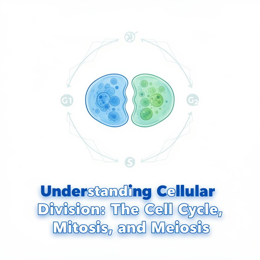
3.1. Phases of the Cell Cycle
The eukaryotic cell cycle consists of five main phases:
- Interphase: The period of growth and DNA replication, comprising G1, S, and G2 phases.
- M Phase (Mitosis): The actual division process, including nuclear division (mitosis) and cytoplasmic division (cytokinesis).
📊 Typical Cultured Human Cell Cycle (approx. 24 hours):
- M phase: ~1 hour
- Interphase: ~23 hours (S phase: ~10-12 hours)
3.2. Interphase: Preparation for Division
Interphase is the portion of the cell cycle between cell divisions.
- G1 Phase (Gap 1):
- Primary growth phase of the cell.
- Major portion of the cell's lifespan for many organisms.
- Cell grows, expresses genes, and differentiates.
- Internal and external conditions are checked.
- Length varies depending on external conditions and extracellular signals.
- S Phase (Synthesis):
- The cell synthesizes a replica of its entire genome (DNA synthesis).
- G2 Phase (Gap 2):
- Second growth phase.
- Preparations for genomic separation are made.
- Mitochondria and other organelles replicate.
- Chromosomes begin to condense.
- Microtubules begin to assemble at a spindle.
- Quality control checks are performed.
3.3. G0 Phase: Quiescence 😴
- If conditions are not suitable during G1, cells may enter the G0 phase, a quiescent state.
- Cells can remain in G0 for days, months, or even years.
- Examples:
- Permanent G0: Many cells in our body, such as neurons and skeletal muscle cells, remain in G0 until the organism dies.
- Temporary G0: Some cells, like damaged liver cells, can re-enter the cell cycle from G0 when needed.
- Repetitive Cycling: Others, like fibroblasts and lymphocytes, enter the cell cycle repeatedly.
4. Eukaryotic Cell Division: Mitosis and Cytokinesis 🔬
The M phase includes mitosis (nuclear division) and cytokinesis (cytoplasmic division). Mitosis is a continuous process but is traditionally divided into four stages.
4.1. Interphase (Prior to Mitosis)
💡 Preparation Stage:
- Chromosomes are decondensed and not individually visible, as they have replicated during the S phase but are not yet condensed.
- The nucleolus is present.
- Two centrosomes have formed by the duplication of a single centrosome. Each centrosome contains a pair of centrioles.
4.2. Stages of Mitosis (M Phase) 🖼️
Mitosis ensures that each daughter cell receives an identical set of chromosomes.
4.2.1. Prophase 🌟
- Nucleolus disappears.
- Mitotic spindle apparatus begins to form: Microtubules extend from the centrosomes. Radial arrays of shorter microtubules around the centrosomes are called asters.
- Centrosomes move away from each other towards opposite poles of the cell.
- Chromatin fibers become more tightly coiled, condensing into discrete chromosomes.
- Each duplicated chromosome appears as two identical sister chromatids joined at their centromere.
- Spindle Apparatus Components:
- Aster microtubules: Important for positioning the spindle apparatus.
- Polar microtubules: Help to "push" the poles away from each other.
- Kinetochore microtubules: Will attach to the kinetochores on the centromeres of chromosomes.
4.2.2. Prometaphase 💥
- Nuclear envelope fragments into vesicles.
- Microtubules invade the nuclear area.
- Chromosomes become even more condensed.
- Each of the two sister chromatids of each chromosome develops a protein structure called a kinetochore at its centromere.
- Kinetochore microtubules attach to the kinetochores, causing the chromosomes to jerk back and forth.
- Polar (non-kinetochore) microtubules interact with those from the opposite pole of the spindle.
4.2.3. Metaphase 📏
- Centrosomes are now at opposite poles of the cell.
- Chromosomes align at the metaphase plate, an imaginary plane equidistant from the two spindle poles.
- The kinetochores of the sister chromatids are attached to kinetochore microtubules coming from opposite poles, ensuring proper tension and alignment.
4.2.4. Anaphase 🏃♀️
- Cohesin proteins are cleaved, leading to the separation of the two sister chromatids.
- Once separated, each chromatid is considered a daughter chromosome.
- Kinetochore microtubules shorten, pulling the newly liberated daughter chromosomes towards opposite ends of the cell.
- Polar microtubules lengthen, causing the cell to elongate.
- By the end of anaphase, the two ends of the cell have equivalent and complete collections of chromosomes.
4.2.5. Telophase ✌️
- Chromosomes reach the poles and begin to decondense.
- Two daughter nuclei form: Nuclear envelopes arise from fragments of the parent cell's nuclear envelope and other portions of the endomembrane system.
- The remaining spindle microtubules depolymerize.
4.3. Cytokinesis: Cytoplasmic Division ✂️
- Cytokinesis typically overlaps with the latter stages of telophase.
- Animal Cells: A cleavage furrow forms. This is a contracting ring of actin and myosin microfilaments that constricts like a drawstring, pinching the cell in two.
- Plant Cells: A cell plate forms in the middle of the cell. Vesicles derived from the Golgi apparatus fuse to form a new cell wall between the two daughter nuclei.
5. Key Outcomes and Importance of Mitosis ✅
- Mitosis ultimately produces two daughter cells that are genetically identical to the mother cell (barring rare mutations).
- Processes requiring mitotic cell division:
- Development of multicellularity: From a single zygote to a complex organism.
- Organismal growth: Increase in size.
- Wound repair: Replacing damaged cells.
- Tissue regeneration: Replacing old or lost cells (e.g., skin cells, blood cells).
Thank you for your patience and attention!