This study material has been compiled from various sources, including a lecture audio transcript, copy-pasted text, and practical task descriptions, to provide a comprehensive overview of the laboratory diagnosis of genitourinary tract infections.
📚 Laboratory Diagnosis of Genitourinary Tract Infections
Introduction
Understanding the precise methods for diagnosing genitourinary tract infections is crucial for effective treatment and patient management. This guide covers both urinary tract infections (UTIs) and sexually transmitted infections (STIs), detailing critical steps from proper specimen collection to the interpretation of laboratory results.
I. Urinary Tract Infections (UTIs)
1. Overview and Types
Urinary Tract Infections (UTIs) are primarily categorized into two types:
- Cystitis: 📚 Infection of the bladder.
- Pyelonephritis: 📚 Infection affecting the renal parenchyma (kidneys).
The urinary tract consists of the kidneys, ureters, bladder, and urethra. While the urethra hosts resident microflora, all areas above it in a healthy person are sterile. Bacterial infection usually occurs via the ascending route from the urethra to the bladder. Females are more prone to UTIs due to a shorter urethra and its proximity to the anus, along with factors like sexual activity, pregnancy, and menopause.
2. Common Etiological Agents
A variety of microorganisms can cause UTIs:
- Enterobacteria:
- E. coli (most common)
- Klebsiella spp.
- Proteus spp.
- Enterobacter spp.
- Other Gram-Negative Bacilli:
- P. aeruginosa
- Acinetobacter spp.
- Alcaligenes spp.
- Gram-Positive Cocci:
- Enterococcus spp.
- Staphylococcus saprophyticus
- Staphylococcus aureus
- Fungi:
- Candida spp.
3. Laboratory Diagnosis of UTIs
a) Proper Collection of Urine Specimens
The accuracy of UTI diagnosis heavily relies on proper specimen collection. Urine in a healthy bladder is sterile, but it can acquire normal flora as it passes through the distal urethra.
- Clean-Catch, Midstream Urine Specimen: ✅ This is the most commonly used method to minimize contamination from indigenous microbes.
- 💡 The first early morning urine sample is preferred as it contains the highest concentration of infecting microorganisms.
- Males: Cleanse the meatus with soap and water, then collect midstream urine in a sterile container.
- Females: Spread the labia and cleanse the vulva before collecting midstream urine.
- Catheterization: Allows collection of bladder urine with less urethral contamination, but carries a risk of introducing microorganisms into the bladder.
- Suprapubic Bladder Aspiration: Ensures contamination-free specimens.
- Specimen Transport: ⚠️ Cultures must be performed within 1 hour after collection or refrigerated (not longer than overnight) because many microorganisms multiply rapidly in urine at room temperature.
b) Direct Microscopic Examination
Microscopic examination of urine provides rapid preliminary information.
- Urine Sediment: A drop of urine sediment (uncentrifuged or centrifuged) examined under a high-dry objective can reveal:
- White Blood Cells (WBCs): Presence of greater than 10 WBCs/µl (known as pyuria 📚) is highly suggestive of bacterial UTIs.
- Epithelial Cells: Squamous epithelial cells of vaginal origin indicate improper collection.
- Bacteria:
- Gram's Stain of Non-Centrifuged Urine: The presence of bacteria is strongly suggestive of 10^5 or more bacteria per milliliter of urine. Observation of at least one bacterium per high-power field is considered equivalent to 10^5 bacteria/ml.
c) Quantitative Cultures
Culture of urine must be performed quantitatively to distinguish contamination from actual infection.
- Method: 1️⃣ Use a bacteriologic loop calibrated to deliver 0.01 ml or 0.001 ml of urine. 2️⃣ Inoculate urine onto agar plates (e.g., 5% Blood Agar Plate (BAP) and MacConkey agar). New chromogenic media (e.g., CPS ID3) are also available for isolation, enumeration, and direct identification of uropathogens. 3️⃣ Count the number of colonies that grow after incubation. 4️⃣ Calculate the number of Colony Forming Units (CFU) per milliliter by multiplying the colony count by 100 (for 0.01 ml loop) or 1000 (for 0.001 ml loop).
- Practical Task: Performance of quantitative urine cultures using a calibrated loop.
d) Interpreting Culture Results
Distinguishing contaminating organisms from etiologically important organisms is crucial.
- Significant Bacteriuria: ✅ Generally accepted as 10^5 or more bacteria (or CFU)/ml of urine, indicative of a UTI, even if the patient is asymptomatic. Recent studies suggest ≥ 10^2 bacteria/ml may also be significant.
- Exceptions:
- Young women with cystitis symptoms: As few as 10^3 CFU/ml of a potential pathogen may be significant.
- Specific patient groups: Less than 10^4 CFU/ml of one potential pathogen may indicate infection in children, males, catheterized patients, or individuals with urinary obstruction.
- Contamination: ⚠️ Growth of less than 10^4 CFU/ml of two or more probable pathogens suggests contamination.
- Practical Task: Interpretation of quantitative urine culture results.
e) Identification of Isolated Microorganisms
Isolated microorganisms are identified using rapid and conventional biochemical tests.
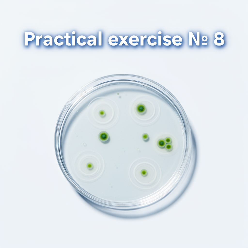
f) Antimicrobial Susceptibility Tests
Antimicrobial susceptibility on clinically relevant bacteria is determined by disk diffusion test.
4. Treatment
Treatment depends on the identified causative agent.
II. Genital and Sexually Transmitted Pathogens (STIs)
1. Overview and Causative Agents
Sexually Transmitted Infections (STIs) are transmitted between humans primarily through sexual behavior, but can also occur via IV drug needles, childbirth, or breastfeeding. They are caused by a diverse range of microorganisms.
| Disease | Causative Agents …