This study material has been compiled from a copy-pasted text and a lecture audio transcript.
📚 Cellular Biology: Structure, Function, and Dynamics
1. Introduction to Cellular Biology and its Evolution
Cellular biology is the branch of biological science dedicated to studying the structure and functions of the cell, the fundamental unit of all living organisms. Our understanding of cells has evolved significantly over centuries, driven by technological advancements and scientific inquiry.
1.1. Historical Milestones 🕰️
- 16th Century: First microscopes invented, enabling initial observations.
- 1655: Robert Hooke discovered the first cells in cork.
- 1674: Anton van Leeuwenhoek observed the first living cells (protozoa, bacteria, blood cells), even describing their movement.
- 19th Century:
- Robert Brown identified the nucleus as a constant component of cells (later refined for eukaryotes).
- The concept of "protoplasm" as living matter containing the nucleus and cytoplasm emerged.
- Modern Cell Theory (Schleiden & Schwann):
- All bodies are composed of cells and substances produced by them.
- Cells possess their own life.
- The life of cells is subordinated to the life of the entire organism.
- Rudolf Virchow: Added the crucial principle: "Omnis cellula e cellula" (all cells arise from pre-existing cells).
- Discovery of cell division (mitosis, meiosis) and chromosomes.
- Virchow also laid the foundation for histopathology by linking diseases to cellular modifications.
- 20th Century:
- Development of advanced techniques: tissue and cell cultures, microsurgery, electron microscopy.
- Electron microscopy revealed ultrastructures like the plasmalemma, ER, peroxisomes, Golgi apparatus, and cytoplasmic differentiations.
- E. Palade's work on ribosomes, mitochondria, cell secretion, and vesicle transport revolutionized the field.
- The "Cell/Molecular Biology Revolution" (1975) integrated techniques like X-ray diffraction and DNA sequencing, shifting focus to molecular-level processes.
1.2. Importance in Medicine 🏥
Knowledge from cell research is paramount as many diseases manifest at the cellular level. Effective treatments rely on understanding these intricate cellular mechanisms.
2. Fundamental Cell Types: Prokaryotic vs. Eukaryotic
Living organisms are broadly categorized into two cell types:
| Feature | Prokaryotic Cells | Eukaryotic Cells | | :------------------ | :---------------------------------------------- | :---------------------------------------------------------------------------- | | Organism Type | Unicellular | Uni- (e.g., protozoa) or Pluricellular (e.g., metazoa) | | Nucleus | Absent (genetic material in nucleoid) | Present (well-defined, membrane-bound) | | Organelles | No membrane-bound organelles | Present (mitochondria, lysosomes, ER, Golgi, peroxisomes, etc.) | | Genetic Material| Single, circular DNA molecule | Multiple, linear DNA molecules (chromosomes) in nucleus; circular in mitochondria | | Cell Division | Direct (amitosis) | Indirect (mitosis, meiosis) | | Cytoskeleton | Simple flagella | Complex (microtubules, filaments) | | Cell Wall | Present (peptidoglycan) | Present in plants (cellulose), fungi; absent in animals | | Size | Typically smaller (1-10 µm) | Typically larger (10-100 µm) | | Evolution | Appeared ~3.5 billion years ago | Appeared ~1.5 billion years ago |
3. Molecular Basis of Cellular Organization
Cells are composed of a precise ratio of chemical substances, primarily water, organic macromolecules, and inorganic ions.
3.1. Chemical Elements ⚛️
Over 60 elements constitute living matter, categorized by abundance:
- Macroelements (2-60%): C, H, O, N. Carbon is key due to its tetravalency, forming diverse macromolecules vital for metabolism.
- Microelements (0.02-0.1%): P, S, Cl (metalloids); Na, K, Ca, Mg (metals). Crucial for structural integrity and physiological functions (e.g., Na+ outside, K+ inside cell).
- Oligoelements (Trace Elements, <0.02%): Fe, I, Co, Zn, Pb. Essential for specific functions (e.g., Fe in hemoglobin for O2 transport, I in thyroid hormones).
3.2. Inorganic Substances 💧
- Water (70%): The "essence of life."
- Properties: Electric dipole (good solvent for polar substances), dissociates into H+ and OH-, forms hydrogen bonds (high caloric capacity, thermal shield, thermoregulation).
- Role: Universal solvent for chemical reactions.
- Transport: Aquaporins facilitate water movement across membranes, implicated in conditions like diabetes.
- Mineral Salts: Present as ions or bound to macromolecules.
- Cations: Na+, K+, Ca2+, Mg2+.
- Anions: PO43-, Cl-, CO32-.
- Role: Influence enzyme activity, membrane permeability, excitability, contractility, osmotic pressure, and acid-base balance. Small variations can be critical.
3.3. Organic Substances 🧬
- Glucids (Carbohydrates):
- Plastic Role: Monosaccharides (ribose, deoxyribose) in nucleic acids.
- Energetic Role: Glucose (main fuel, high energy release, ATP synthesis via glycolysis); Glycogen (storage form of glucose in liver/muscles).
- Structural: Mucopolysaccharides (e.g., hyaluronic acid) in connective tissue.
- Lipids:
- Plastic Role: Cell membranes (phospholipids, glycolipids).
- Energetic Role: Highest energy value (fatty acids, triglycerides).
- Regulatory Role: Hormones, vitamins.
- Membrane Components: Amphiphilic nature (hydrophilic head, hydrophobic tail) forms lipid bilayers. Cholesterol modulates fluidity.
- Proteins (15%): Highly diverse, performing myriad functions.
- Roles: Structural support, storage, transport (e.g., hemoglobin), muscle contraction (myosin), resistance/elasticity, regulation (hormones), catalysis (enzymes), immune defense, receptors, osmotic pressure/pH maintenance.
- Specificity: Unique amino acid sequences and 3D structures allow precise binding (e.g., enzyme-substrate).
- Nucleic Acids (DNA, RNA):
- Structure: Macromolecules of nucleotides (nitrogenous base, pentose, phosphate).
- DNA: Stores and transmits genetic information (heredity, variability). Located in nucleus (chromosomes) and mitochondria.
- RNA: Involved in protein synthesis. Located in nucleus (nucleolus) and cytoplasm (rRNA, tRNA, mRNA).
4. Cellular Dynamics and Structures
4.1. Cytoplasmic Matrix & Cytoskeleton 🏗️
The cytoplasm, excluding organelles, is the cytosol, a homogenous system where macromolecules are dissolved. It exhibits colloidal phenomena like diffusion and sol-gel transitions. The cytoskeleton is a dynamic 3D network of protein filaments (actin, microtubules, intermediate filaments) providing structural support, enabling cell movement, and organizing organelles.
- Actin Filaments (Microfilaments): Smallest (5 nm), involved in cell shape, cortical network, contractile rings, and microvilli movement.
- Muscle Cells: Form "thin" filaments, interacting with myosin for contraction.
- Myosin Filaments: Motor proteins with ATPase activity, interacting with actin for muscle contraction and amoeboid movement.
- Intermediate Filaments: Medium size (10 nm), very stable, provide mechanical strength (e.g., keratin in epithelia, lamins in nuclear envelope).
- Microtubules: Largest (25 nm), made of tubulin. Dynamic structures involved in cell shape, organelle transport, cilia/flagella movement, and cell division (mitotic spindle).
- Medical Importance: Affected by drugs like colchicine (inhibits polymerization, used in gout) and taxol (stabilizes, used in cancer).
4.2. Cell Motility 🏃♀️
Achieved by motor proteins (e.g., myosin, kinesin, dynein) using ATP to change protein conformation and generate movement.
- Actin-Myosin System: Muscle contraction, amoeboid locomotion (e.g., neutrophils, fibroblasts), microvilli movements.
- Microtubule-Dynein System: Axonal transport (anterograde by kinesin, retrograde by dynein), movements of cilia and flagella.
- Cilia/Flagella: Permanent extensions with an internal axoneme (9+2 microtubule doublets) for coordinated movement. Defects can cause respiratory infections and sterility (e.g., Kartagener syndrome).
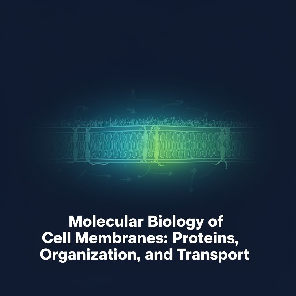
4.3. Cell Membranes: Structure, Function, Transport 🛡️
Two-dimensional structures of proteins and lipids that separate cellular compartments and the cell from its environment.
- Functions: Barrier, compartmentalization, metabolic functions, information flow, signal transduction, immunity.
- Composition: Lipids (phospholipids, glycolipids, cholesterol) and proteins. Protein:lipid ratio varies with function.
- Fluid Mosaic Model (Singer & Nicholson): Lipid bilayer is fluid, allowing proteins to move. Glycoproteins and glycolipids have extracellular sugar groups.
- Transport Across Membranes:
- Passive Transport: No energy, down electrochemical gradient.
- Simple Diffusion: Liposoluble substances directly through bilayer.
- Facilitated Diffusion: Via transporter proteins (saturable).
- Channel Proteins: Rapid transport (e.g., aquaporins, gated channels).
- Active Transport: Requires ATP, against electrochemical gradient.
- Ionic Pumps: Ca2+ pumps, Na+-K+ pump (maintains membrane potential, cell volume).
- ATP-binding Cassette (ABC) Transporters: Transport various substances (e.g., MDRP in detoxification, CFTR for Cl-).
- Coupled Transport: Uses ionic gradients (e.g., glucose-Na+ symporter).
- Macromolecule Transport (Vesicular):
- Exocytosis: Release of substances (secretion products, neurotransmitters).
- Endocytosis: Uptake of substances.
- Phagocytosis: Uptake of solid material (e.g., bacteria by macrophages).
- Pinocytosis: Uptake of liquids (receptor-dependent or independent).
- Transcytosis: Transcellular transport across the cell.
- Passive Transport: No energy, down electrochemical gradient.
4.4. Cell Junctions 🤝
Stable structures mediating intercellular communication and adhesion.
- Adherens Junctions (Desmosomes): Provide mechanical strength (e.g., belt desmosomes, spot desmosomes, hemidesmosomes).
- Tight Junctions: Prevent leakage between cells, forming a barrier (e.g., blood-brain barrier).
- Gap Junctions: Direct communication channels between cells via connexons, allowing passage of ions and small molecules.
4.5. Cell Recognition, Aging, and Death 💀
- Cell Recognition: Cells interact specifically (e.g., immune response, sperm-egg binding, contact inhibition in cultures). Malignant cells often lose contact inhibition.
- Cell Aging: Characterized by decreased metabolic activity, changes in nuclear and cytoplasmic ratios, and accumulation of pigments/lipids.
- Cell Death:
- Necrosis: Accidental, uncontrolled cell death due to injury (e.g., anoxia, burns). Leads to inflammation.
- Apoptosis: Programmed, physiological cell death. Involves specific proteases (caspases), DNA degradation, and phagocytosis by neighboring cells without inflammation. Crucial for development and tissue turnover.
5. Nucleus & Chromosomes
The nucleus is the control center of eukaryotic cells, storing most genetic material and coordinating cellular functions.
5.1. Nucleus Structure (Interphase) ⚛️
- Nuclear Envelope: Double membrane with nuclear pores, continuous with RER. Nuclear lamina (intermediate filaments) provides structural support.
- Nuclear Pores: Complex structures regulating transport between nucleus and cytoplasm (e.g., import of polymerases, export of RNA).
- Chromatin: DNA and histones.
- Euchromatin: Relaxed, metabolically active, transcribed.
- Heterochromatin: Condensed, metabolically inactive (e.g., Barr body, inactive X chromosome).
- Nucleolus: Site of ribosome biogenesis (rRNA synthesis and assembly with ribosomal proteins).
- Karyoplasm: Nuclear matrix, containing proteins, enzymes, nucleotides, and ions.
5.2. Eukaryotic Chromosomes 🧬
Main structures of heredity, carrying genetic information in DNA sequences.
- Functions: Storage, transmission (replication), and expression (transcription, translation) of genetic information.
- Morphology: Studied during anaphase, characterized by centromere (primary constriction), secondary constrictions, and satellites.
- Karyotype: Number and morphology of chromosomes in metaphase. Human somatic cells are diploid (2n=46), gametes are haploid (n=23).
- Autosomes: 22 pairs.
- Gonosomes: 1 pair (XX for female, XY for male).
- Abnormalities: Numeric (e.g., Trisomy 21/Down Syndrome, Klinefelter, Turner) or structural (deletions, translocations).
- Chromatin Organization: DNA is wrapped around histone octamers to form nucleosomes, which then coil into chromatin filaments and thicker fibers, ultimately forming chromosomes.
5.3. Cell Division ➗
- Cell Cycle: G1 (growth), S (DNA replication), G2 (preparation for division), M (mitosis/meiosis). Regulated by cyclins and cyclin-dependent kinases (Cdks).
- Mitosis: Indirect division of diploid cells, producing two identical diploid daughter cells.
- Phases: Prophase (chromatin condensation, mitotic spindle formation), Prometaphase (nuclear envelope disassembly, chromosome attachment to spindle), Metaphase (chromosomes align at equatorial plate), Anaphase (sister chromatids separate), Telophase (chromosomes decondense, nuclear envelope reforms), Cytokinesis (cytoplasmic division).
- Meiosis: Indirect division generating haploid germ cells (gametes) from diploid cells, involving one DNA replication and two divisions (Meiosis I and Meiosis II).
- Key Event (Prophase I): Crossing over between homologous chromosomes, leading to genetic recombination and increased variability.
6. Endoplasmic Reticulum (ER)
A network of intracellular membranes continuous with the outer nuclear membrane.
- Rough ER (RER): Studded with ribosomes.
- Function: Synthesis of proteins for secretion, insertion into membranes, or delivery to organelles like Golgi, lysosomes. Involves "vectorial transfer" of polypeptide chains into the ER lumen.
- Smooth ER (SER): Lacks ribosomes.
- Function: Lipid synthesis (e.g., triglycerides, phospholipids), detoxification of drugs/compounds (especially in liver), glycogen degradation.
- Common Functions: Phospholipid synthesis, membrane biogenesis, intracellular transport, mechanical support, Ca2+ reservoir.
7. Golgi Apparatus (GA) & Cell Secretion
A system of flattened sacs (cisternae) and vesicles, typically near the nucleus.
- Structure: Cis (convex, receiving side from ER), medial, and Trans (concave, shipping side) faces.
- Functions:
- Processing & Packaging: Biochemical modification (proteolysis, terminal glycosylation, sulfation), concentration, sorting, and packaging of proteins and lipids from the ER into secretion vesicles.
- Biogenesis: Formation of lysosomes and new membranes.
- Coordination: Regulates membrane traffic within the cell.
- Cell Secretion: A common activity in eukaryotic cells (e.g., digestive enzymes, hormones). Involves RER (synthesis), ER (transport), GA (processing), and secretion vesicles (transport to membrane for exocytosis).
8. Lysosomes
Membrane-bound organelles containing hydrolytic enzymes active at acidic pH (4.5-5).
- Enzymatic Content: Over 40 enzymes capable of digesting all cellular compounds (DNA, RNA, proteins, lipids, carbohydrates).
- Latency: Enzymes are inactive when the lysosomal membrane is intact.
- Biogenesis: Lysosomal enzymes are synthesized in RER, tagged with mannose-6-phosphate in the Golgi, and then packaged into vesicles that mature into lysosomes.
- Functions (Intracellular Digestion):
- Heterophagy: Digestion of foreign materials taken up by endocytosis (phagocytosis, pinocytosis).
- Autophagy: Digestion of old or damaged cellular components (e.g., organelles).
- Crinophagy: Control of secretion product amount by digesting excess.
- Medical Implications: Essential for defense, but defects can lead to lysosomal storage diseases (e.g., Gaucher, Tay-Sachs) or contribute to inflammation (e.g., rheumatism).
9. Peroxisomes
Small, spherical organelles involved in cellular oxidation.
- Enzymes: Contain oxidases (using O2 to generate H2O2) and catalase (detoxifies H2O2).
- Functions: Oxidation of fatty acids, detoxification of alcohols/phenols, synthesis of plasmalogens.
- Medical Implications: Peroxisomal genetic diseases (e.g., Zellweger syndrome, adrenoleukodystrophy) result from defects in their biogenesis or enzyme function.
10. Mitochondria
The "powerhouses" of the cell, responsible for ATP production via oxidative phosphorylation.
- Structure: Double membrane (outer permeable, inner folded into cristae and largely impermeable). Contains its own circular DNA, ribosomes, and matrix.
- Metabolic Processes:
- Citric Acid Cycle (Krebs Cycle): In matrix, produces NADH and FADH2.
- Oxidative Phosphorylation: In inner membrane, electrons from NADH/FADH2 pass through an electron transport chain, creating a proton gradient. This gradient drives ATP synthase to produce ATP (chemiosmotic theory).
- Biogenesis: Arise from pre-existing mitochondria, with some components synthesized internally and others imported from the cytosol.
- Medical Implications:
- Genetic Mitochondrial Diseases: Mutations in mtDNA or nuclear genes affecting mitochondrial function (e.g., Leber's disease, MERF syndrome). Tissues with high ATP demand (muscles, CNS) are most affected.
- Involved in various diseases (cancer, cardiovascular) and apoptosis.
11. Extracellular Matrix (ECM) & Cell Adhesion
A network of macromolecules occupying the intercellular space, providing structural and biochemical support.
- Composition: Fibrous proteins (collagen, elastin, fibronectin, laminin) embedded in a hydrated polysaccharide gel (glycosaminoglycans, proteoglycans).
- Functions: Tissue structure, lubrication, elasticity, cell adhesion, influences cell shape/migration/development, metabolic role, immunity.
- Cell Adhesion Molecules: Specific proteins mediating cell-cell or cell-matrix interactions (e.g., selectins, immunoglobulins, cadherins, integrins).
- Basal Lamina: Specialized ECM under epithelial cells, acting as a semi-permeable filter and barrier.
12. Central Dogma of Molecular Biology & DNA Replication
The fundamental principle describing the flow of genetic information within a cell: DNA → RNA → Protein.
- Genetic Material: DNA (in chromosomes and mitochondria) stores information in nucleotide sequences.
- DNA Replication: The process by which DNA is duplicated.
- Semiconservative: Each new DNA molecule consists of one original strand and one newly synthesized strand.
- Mechanism: DNA unwinds, and DNA polymerase synthesizes new strands (leading and lagging strands with Okazaki fragments).
- Medical Implications:
- Mutations: Errors during replication or damage from chemical/physical agents (UV, ionizing radiation) can lead to mutations. Cells have repair mechanisms.
- Inhibitors: Drugs targeting DNA synthesis (antibiotics, anticancer drugs) exploit differences between host and pathogen/cancer cell replication.